
Pumping strength
How strongly the heart pushes out blood with each beat. Your report may give this as EF (ejection fraction), a percentage.
An ultrasound of the heart by visiting cardiologists from Rhythm Heart Institute, Vadodara: every Thursday, 5 to 6 PM, by appointment. Report the same evening.
Visiting cardiologists from Rhythm Heart Institute, Vadodara
A 2D Echo is an ultrasound of your heart. It shows your heart beating on a screen, so that a cardiologist can see how well it pumps and how its valves are working.
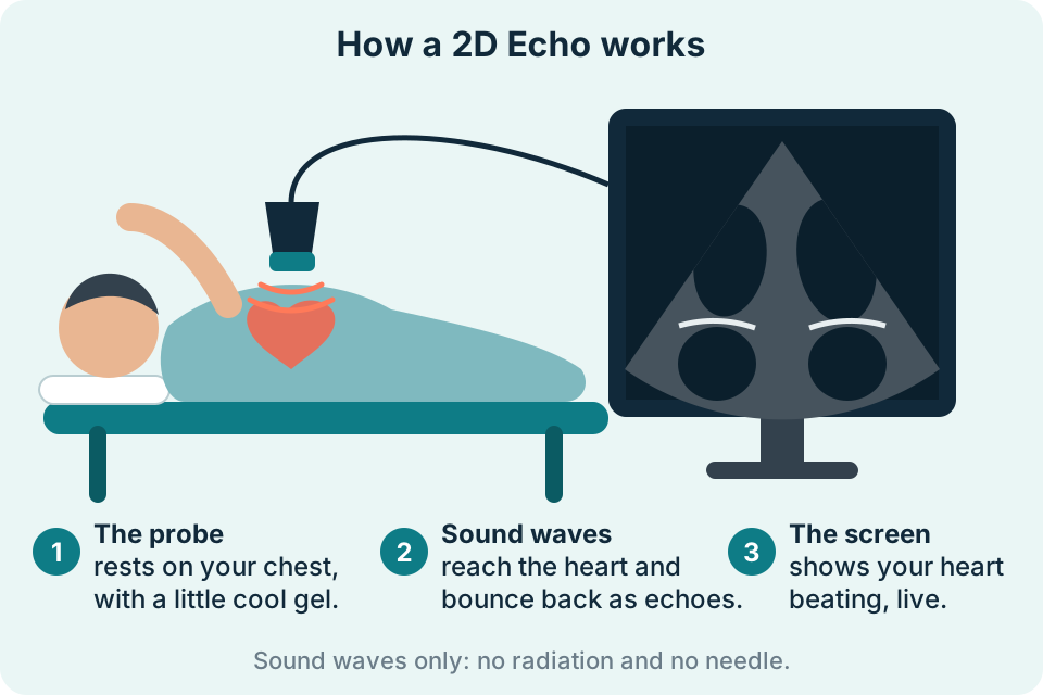
A 2D Echo is a safe, painless test that lets a heart specialist watch your heart while it beats. It works like a pregnancy ultrasound, but the probe is placed on the chest. There is no radiation and no injection.
Why “2D”? The machine shows thin, flat slices of the heart as moving pictures. By looking from different positions on the chest, the cardiologist can see every part of the heart.
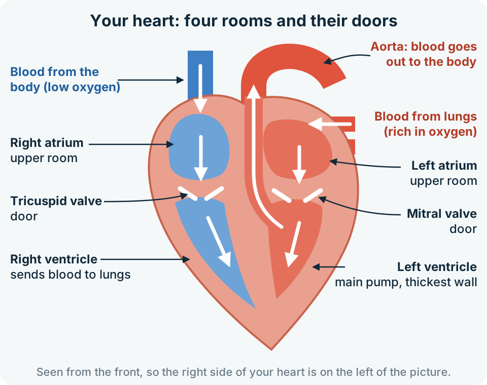
Think of your heart as a small house with four rooms. The two upper rooms (atria) receive blood. The two lower rooms (ventricles) pump it out. Four doors, called valves, open to let blood through and shut so that it does not flow back.
The right side sends blood to the lungs to pick up oxygen. The left side, the strongest part, then pumps that blood to the whole body.
You lie on a couch, usually turned towards your left side, because this brings the heart closer to the chest wall. The cardiologist puts a little gel on your chest and moves a small probe over it. The probe sends out sound waves that are too high for you to hear. They bounce back from the heart as echoes, and the machine turns these echoes into a moving picture.
The test shows how well your heart is working, not only how it looks.
Visiting cardiologists from Rhythm Heart Institute, Vadodara, a well-known heart hospital with multiple branches, come to our centre every Thursday from 5 to 6 PM. You get a heart specialist’s test here in Halol, without travelling to Vadodara.
The test is by appointment. Please call 84475 86847 or 82385 43753 to book your Thursday slot. For a scan of an unborn baby’s heart, see Fetal Medicine.
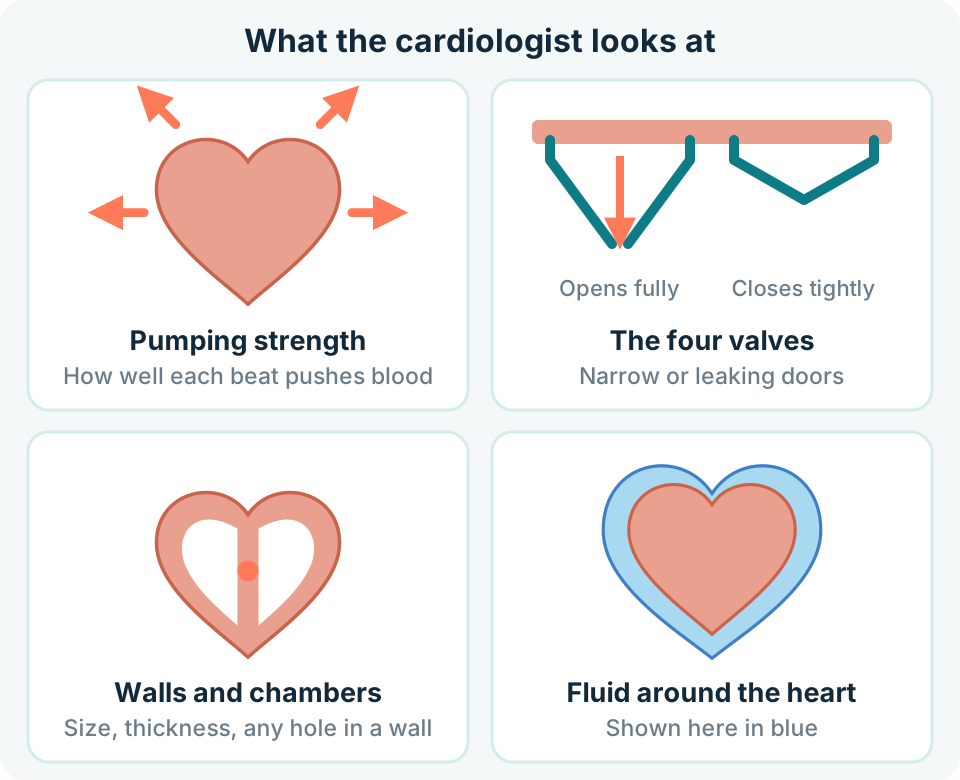
Your report may mention some of these. Your doctor will explain what the results mean for you.
How strongly the heart pushes out blood with each beat. Your report may give this as EF (ejection fraction), a percentage.
Whether each valve opens fully and closes tightly. A valve can become narrow (stenosis) or leaky (regurgitation).
The size of each chamber and the thickness of the heart muscle. A part of the wall that moves poorly can be a sign of reduced blood supply.
Openings in the walls between the chambers (ASD or VSD) and other heart conditions present from birth.
Extra fluid in the thin sac around the heart, known as pericardial effusion.
An estimate of the pressure in the blood vessels of the lungs.
Call 84475 86847 or 82385 43753 to book a Thursday appointment between 5 and 6 PM.
Please come on time and bring your doctor's slip and old reports. You will be asked to uncover your chest and lie down, usually turned towards your left side.
The cardiologist moves a small probe with gel over your chest. Just lie still and breathe normally. You may be asked to hold your breath for a moment.
You get your report the same evening. Show it to the doctor who advised the test.
No fasting is needed. Eat and drink as usual.
Take your usual medicines unless your doctor has told you otherwise.
Your doctor's slip, old echo and ECG reports, and a list of your medicines.
Loose, two-piece clothes make it easier to reach the chest.
A parent stays with the child. A favourite toy helps a child stay calm and still.
Your doctor may give you different instructions for your test; follow those.
Yes. A 2D Echo uses sound waves, not X-rays, so there is no radiation. It can be repeated whenever your doctor needs it.
No. The gel feels cool and you may feel light pressure from the probe. No needle or injection is used in a routine 2D Echo.
It usually takes about 15 to 30 minutes.
An ECG records the heart's electrical signals. A 2D Echo shows the heart's structure and how it moves. Angiography shows the arteries of the heart directly. A 2D Echo cannot rule out blockages in these arteries, so your doctor may advise other tests if blockages are suspected.
Yes. It uses no radiation, and it is safe if your doctor advises it to check your heart. It does not check the baby's heart; that is a separate fetal echocardiography scan, described on our Fetal Medicine page.
Yes. We do the test for both adults and children. It is painless, and a parent stays with the child.
You get your report the same evening. Please show it to the doctor who advised the test.
Visiting cardiologists from Rhythm Heart Institute, Vadodara, come to our centre every Thursday from 5 to 6 PM. The test is by appointment, so please call 84475 86847 or 82385 43753 to book. If Thursday is a public holiday, please check when you call.
We’re open Mon – Sat: 9:00 AM – 7:00 PM | Sun: By Appointment Only | 2D Echo: Thu 5–6 PM | MRI / CT / XRAY / OPG / Pathology: 24×7 available — walk in or call ahead.