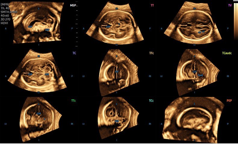
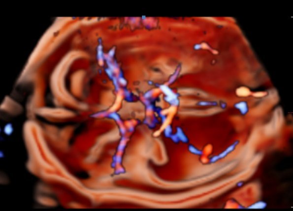
Our fetal scans are done on the Samsung HERA W10, a premium ultrasound machine dedicated to obstetrics and fetal medicine.
At a glance
- Machine
- Samsung HERA W10, used only for pregnancy and fetal scans
- Open
- Mon–Sat 9 AM–7 PM
- Appointment
- Needed for anomaly scans and 3D/4D scans
- Scan time
- 20 to 45 minutes for most scans; the anomaly scan and fetal echo take longer
- 3D/4D face pictures
- Best at about 26 to 30 weeks
- Sex of the baby
- Not told: sex determination is prohibited under the PCPNDT Act
Reviewed by Dr Abhishek Agarwal, Consultant Radiologist & Director ·
Our fetal scans are done on the Samsung HERA W10, a premium ultrasound machine built for obstetrics and fetal medicine. Its fine image detail and dedicated fetal tools help us look at the baby's brain, heart, spine and growth more clearly and more consistently.
What the HERA W10 adds
The 5D tools take a single 3D sweep and automatically find the standard planes and measurements, so results depend less on the angle of the probe and the exam is quicker.
- 5D NT – finds the true mid-line view of the baby in the first-trimester scan and measures the nuchal translucency (NT) at its widest point.
- 5D CNS+ – shows the standard planes of the fetal brain and takes the key brain measurements from one volume.
- 5D Heart Color and HeartAssist – 5D Heart Color displays nine standard fetal-heart views, with colour flow, from one heart volume; HeartAssist helps with heart views and measurements.
- 5D Limb Vol. – measures the volume of the fetal thigh or arm, which helps estimate the baby's weight.
- 5D Follicle – counts and measures the ovarian follicles automatically from one 3D sweep, for monitoring during fertility treatment.
- STIC 4D fetal echo – records one full heartbeat cycle in 3D, so the beating heart can be reviewed from any angle.
- MV-Flow and LumiFlow – MV-Flow shows slow flow in very small vessels, such as in the placenta and the fetal brain; LumiFlow shows blood flow with a 3D-like depth.
- CrystalVue and CrystalVue Flow – 3D renderings that show inside and outside structures together, and blood vessels in 3D.
- Realistic 3D/4D imaging of the face, hands, feet and spine.
4D fetal heart (STIC)
Two short STIC clips from our HERA W10, showing blood flowing through the beating fetal heart:
Dr Rohit Sanghani visits our centre regularly and offers specialised expert services for fetal imaging and interventions. He is a fetal medicine specialist, trained at the renowned Adi Imaging Centre, Bangalore.
When a fetal MRI is needed, it is done on our 1.5 Tesla MRI and reported by Dr Sachin G Shatagar, a radiologist with a fellowship in fetal medicine.
See our images
Which scan, which week
Your doctor decides which scans you need and when. This is the usual pattern:
| Weeks of pregnancy | Scan | What it checks |
|---|---|---|
| 6 to 10 | Early pregnancy (dating) scan | That the pregnancy is in the womb, the heartbeat, the number of babies and the due date |
| 11 to 13 weeks 6 days | NT scan | The nuchal translucency and the baby’s early anatomy; with a blood test, screening for conditions such as Down syndrome |
| 18 to 22 | Anomaly scan (TIFFA) | A detailed check of the baby’s brain, face, spine, heart, abdomen, kidneys and limbs |
| Usually 18 to 24, when advised | Fetal echo | A detailed check of the baby’s heart |
| 26 to 30 (optional) | 3D/4D scan | Pictures of the baby’s face; the best pictures usually come in these weeks |
| 28 onwards | Growth scan and Doppler | The baby’s growth and weight, the fluid, the placenta and blood flow |
Anomaly scans and 3D/4D scans need an appointment: call 84475 86847 or 82385 43753.
▸ Fetal Medicine gallery – fetal heart, brain, growth and Doppler images and STIC clips from our HERA W10.
▸ 3D/4D ultrasound gallery and our Fetal 3D/4D Gallery page.
▸ Meet our HERA W10 – a short read on what the machine does · Fetal medicine FAQs
Sex determination of the fetus is not done at this centre. It is prohibited and punishable under the PCPNDT Act, 1994.
Key features
Watch the video
Frequently asked questions
The NT (nuchal translucency) scan is done between 11 weeks and 13 weeks 6 days of pregnancy. It is usually combined with a blood test (double marker) to estimate the chance of Down syndrome and other chromosomal conditions, and it also checks the baby's early anatomy. On our HERA W10, 5D NT helps find the correct view and measure the NT accurately.
The anomaly scan, also called TIFFA (Targeted Imaging For Fetal Anomalies) or level II scan, is a detailed check of the baby's brain, face, spine, heart, abdomen, kidneys and limbs. It is best done between 18 and 22 weeks of pregnancy.
Fetal echocardiography is a detailed ultrasound of the baby's heart, usually done at about 18 to 24 weeks. Your doctor may advise it if you have diabetes, a family history of heart disease from birth, an IVF pregnancy, a raised NT, or if a routine scan raised a question about the heart. We use STIC 4D and 5D Heart Color on the HERA W10 to review the beating heart from every angle.
The 5D tools on our Samsung HERA W10 (5D NT, 5D CNS+, 5D Heart Color, 5D Limb Vol. and 5D Follicle) take a single 3D sweep and automatically find the standard views and measurements. This makes results more consistent and the exam quicker. For you, the scan feels exactly the same as a normal ultrasound.
The best pictures usually come between about 26 and 30 weeks. Good pictures also depend on the baby's position and the amount of fluid in front of the face, so sometimes we ask you to walk for a while and try again.
Yes. 3D and 4D scans use the same sound waves as a normal pregnancy ultrasound. There is no radiation. We follow standard safety settings and scan only for as long as needed.
Most pregnancy scans take about 20 to 45 minutes. The anomaly scan and fetal echo take longer. If the baby is lying in a difficult position, we may ask you to walk or change position and then continue the scan.
No. Telling or hinting at the sex of the fetus is prohibited and punishable under the PCPNDT Act, 1994. It is not done at our centre.
Gallery