
Case list
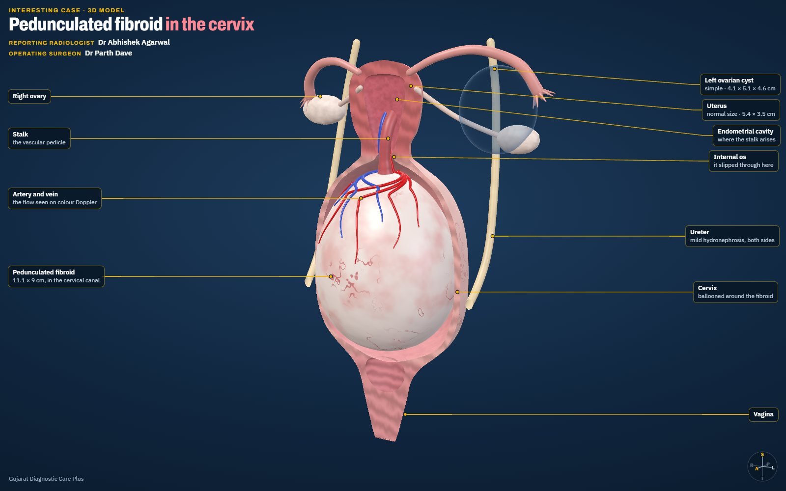
Pedunculated Fibroid in the Cervix: Trace the Stalk
Cervical mass? Trace the stalk.
A 38-year-old woman with a Foley catheter in place. Ultrasound showed a 111 × 90 mm mass filling the cervical canal, with mild hydronephrosis on both sides. Colour Doppler traced its stalk to the endometrium, and surgery confirmed a pedunculated fibroid. An interactive 3D model shows how it hangs on its stalk.
View case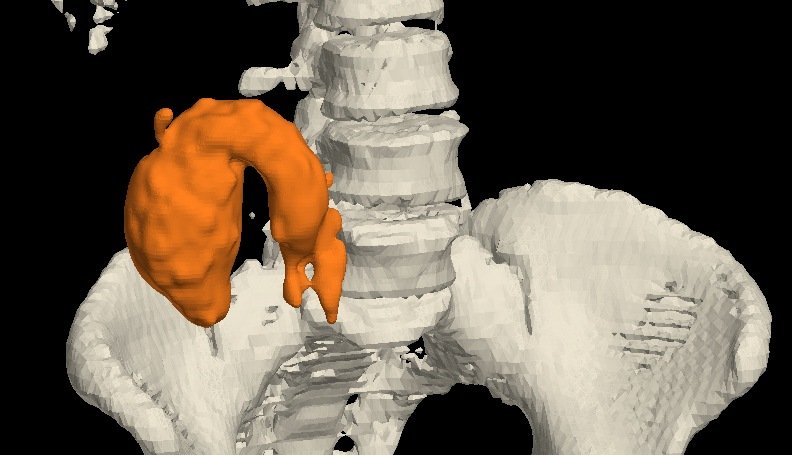
Appendiceal Mucocele: A Fluid-Filled Sausage in the Right Iliac Fossa
Fluid-filled sausage? Think mucocele.
A 50-year-old woman. Contrast CT showed a fluid-filled appendix, 8.9 cm long and 3.8 × 3.9 cm wide, reaching the subhepatic region, with a normal base…
View case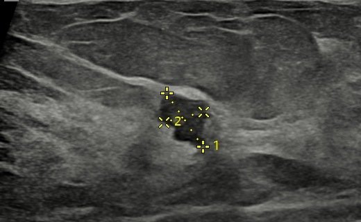
Carcinoma in a Dense Breast: Subtle Distortion, Chased to a Diagnosis
Subtle distortion? Chase it.
A 37-year-old woman with vague left breast pain for a few weeks. Mammography showed heterogeneously dense breasts with mild architectural distortion a…
View case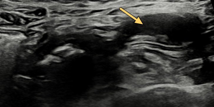
A Twist in the Tail: Meckel's Diverticulum Torsion
Pain and vomiting? Find the blind-ending loop.
A 21-year-old woman with acute abdominal pain and vomiting. Ultrasound showed a dilated pelvic loop with absent flows and an adjacent blind-ending tub…
View case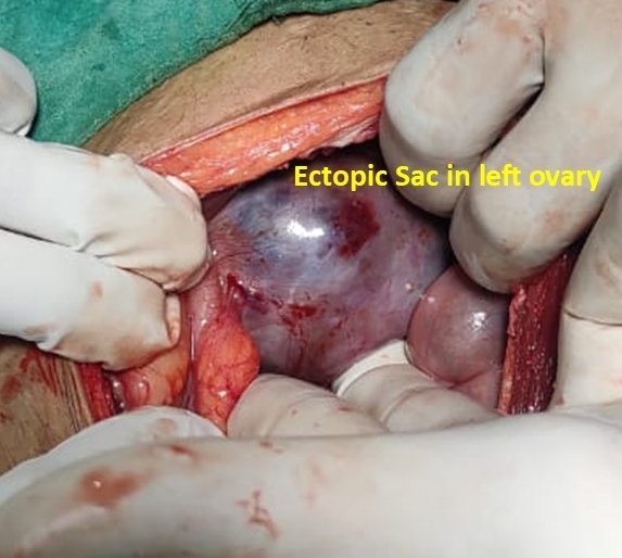
Ovarian Ectopic Pregnancy on Ultrasound
An ectopic in the ovary itself: rare, under 3% of ectopics.
Ultrasound showed a large 6 cm gestational sac in the left adnexa, close to the left ovary, with a fetal pole of 7 weeks 5 days and no cardiac activit…
View case
Torsion of a Meckel's Diverticulum with Gangrene
A dilated pelvic loop and a twisted mesentery: think Meckel's.
Ultrasound showed a focal dilated loop in the pelvis with calcific densities, a twisted mesentery and vessels, and dilated small-bowel loops around it…
View case
Ovary in the Inguinal Canal: Canal of Nuck Hernia
Inguinal swelling in a young woman? The ovary may be inside.
A 20-year-old woman with left inguinal pain and swelling. Ultrasound showed a non-reducible left inguinal hernia containing the left ovary, through a …
View case
A Lump in One Breast, Calcifications in the Other: Why Mammography Matters
A lump in the left breast, and suspicious calcifications in the right.
An 85-year-old woman with a left breast lump. Bilateral mammosonography showed a spiculated malignant-looking lesion with nipple retraction in the lef…
View case
Small VSD on Fetal Echocardiography
A small hole, caught before birth: to-and-fro flow across the septum.
Fetal echocardiography at 25 weeks 4 days showed a small ventricular septal defect, with to-and-fro flow through the defect on colour Doppler.
View case
Endometrial Polyp on 3D Ultrasound: Adding Colours
One polyp, six views: 3D ultrasound of the uterine cavity.
A rounded endometrial polyp seen in the coronal plane of a 3D ultrasound of the uterus, then rendered in different colour and light modes so that the …
View case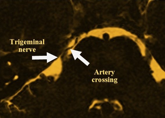
Trigeminal Neuralgia: An Artery Crossing the Nerve on CISS MRI
Electric bursts of facial pain? Look for an artery on the nerve.
Sudden, severe paroxysms of right-sided facial pain lasting seconds to minutes. A high-resolution CISS MRI sequence showed an artery crossing over the…
View case
Non-Contrast MR Renal Angiography
The renal arteries on MRI, with no contrast at all.
A non-contrast MR angiogram shows the abdominal aorta and both renal arteries without any contrast injection: a good way to assess renal artery stenos…
View case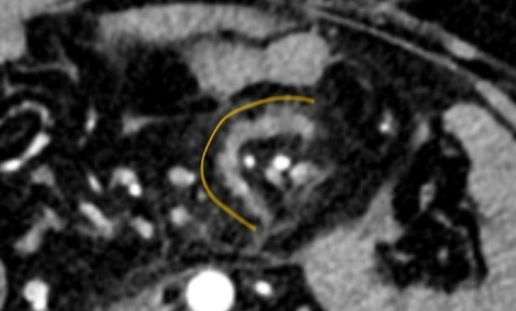
A Tear in the Fan: Traumatic Mesenteric Tear
Blood in the belly, organs intact? Look at the fan.
A 34-year-old man after blunt abdominal injury. Contrast CT showed haemoperitoneum, a C-shaped mesenteric density and a partial SMV thrombus. Laparosc…
View case
Pouches in a Spin: Jejunal Diverticula and Volvulus
No malrotation, yet a whirl? Hunt for the lead point.
A 75-year-old man with pain abdomen and vomiting. Doppler showed a whirlpool of mesenteric vessels; CECT confirmed small-bowel obstruction with an ant…
View case
Uterine & Bowel Perforation after a Failed IUCD Removal
Threads not seen? Scan first.
IUCD threads were not seen and a blind instrument search failed. Next day, ultrasound showed a fundal defect, free fluid and peritoneal thickening. At…
View case
Ileal Diverticulum with a Band: Small-Bowel Obstruction
Small-bowel obstruction with an inflamed pouch? Think diverticulum.
A 58-year-old man with abdominal pain and distension for 2 days. CT showed a diverticulum from a proximal ileal loop with surrounding inflammatory cha…
View case
Hypertrophic Pyloric Stenosis in a 6-Week-Old
Vomiting after every feed? Measure the pylorus.
A 1.5-month-old baby had persistent vomiting after each feed and was not gaining weight. Ultrasound showed the typical pyloric wall thickening of hype…
View case
Jogger's Knee Pain: A Stress Fracture, Not an Osteoid Osteoma
A dense spot on the X-ray after starting to jog? Get the MRI.
A 30-year-old woman had knee pain and difficulty walking for a week after she started jogging. The X-ray raised the question of an osteoid osteoma in …
View case
Peptic Perforation Seen on Ultrasound
Severe epigastric pain? Ultrasound may show the hole.
A 50-year-old man with severe acute epigastric pain. Ultrasound showed echogenic free fluid and a 7 mm defect in the anterior wall of the antropyloric…
View case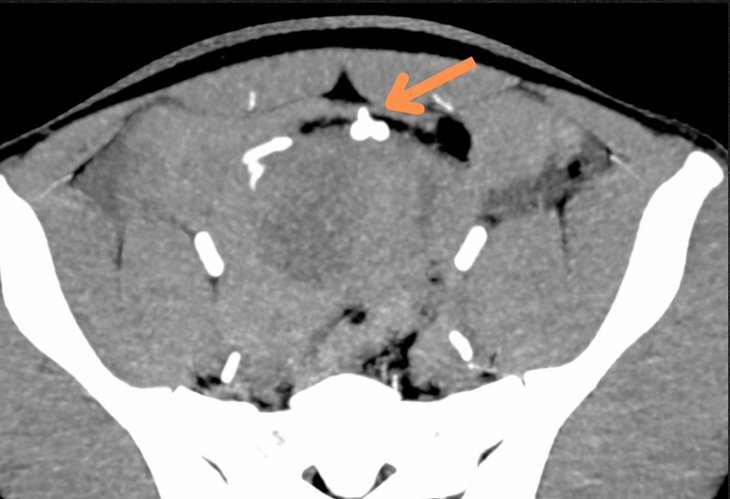
Active Mesenteric Bleed on CT Angiography
Blunt trauma with hypotension? CT angiography finds the bleeder.
A young patient with blunt abdominal trauma and hypotension. CT angiography showed an arterial leak in the pelvis from a mesenteric branch, with activ…
View case
A Large Left Parietal Arteriovenous Malformation
Seizures with arm weakness? Look for the nidus.
A 50-year-old man with seizures and right upper-limb weakness. MRI brain showed a large arteriovenous malformation in the left parietal lobe: a diffus…
View case
The Boomerang Sign: A Splenial Lesion in Hypoglycaemia
Bright splenium after hypoglycaemia? Usually reversible.
A 70-year-old woman suddenly lost consciousness, and lab reports showed hypoglycaemia. MRI brain, done to rule out cerebral injury, showed increased s…
View case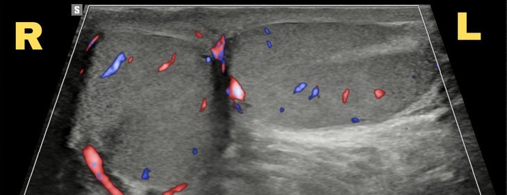
Bell Clapper Deformity: A Testis Lying Sideways
Testicular pain that eased on its own? Check how the testis lies.
Sudden severe pain in the left testis that eased after a few hours. Scrotal ultrasound showed the left testis lying transversely in the sac, the right…
View case
Thyroglossal Fistula: Tracing the Tract on CT Sinogram and MRI
A neck sinus that keeps discharging? Trace the whole tract.
A long-standing sinus in the midline of the neck with occasional pus discharge. A CT sinogram traced the fistula up to the foramen caecum at the base …
View case
Case 1: Ruptured Appendicitis on X-Ray and Ultrasound
An interesting case where ultrasound and X-ray together confirmed acute appendicitis with rupture — later confirmed at surgery.
View caseNo cases match your search.
More from Dr Abhishek
Need a scan or an appointment?
We’re open Mon – Sat: 9:00 AM – 7:00 PM | Sun: By Appointment Only | 2D Echo: Thu 5–6 PM | MRI / CT / XRAY / OPG / Pathology: 24×7 available — walk in or call ahead.