
Latest posts
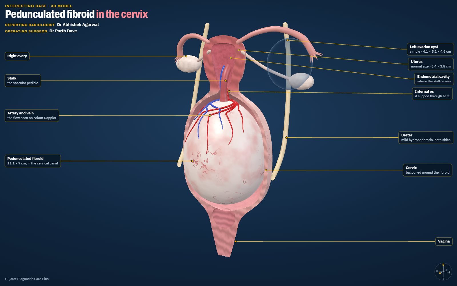
Pedunculated Fibroid in the Cervix: Trace the Stalk
Cervical mass? Trace the stalk.
A 38-year-old woman with a Foley catheter in place. Ultrasound showed a 111 × 90 mm mass filling the cervical canal, with mild hydronephrosis on both sides. Colour Doppler traced its stalk to the endometrium, and surgery confirmed a pedunculated fibroid. An interactive 3D model shows how it hangs on its stalk.
View case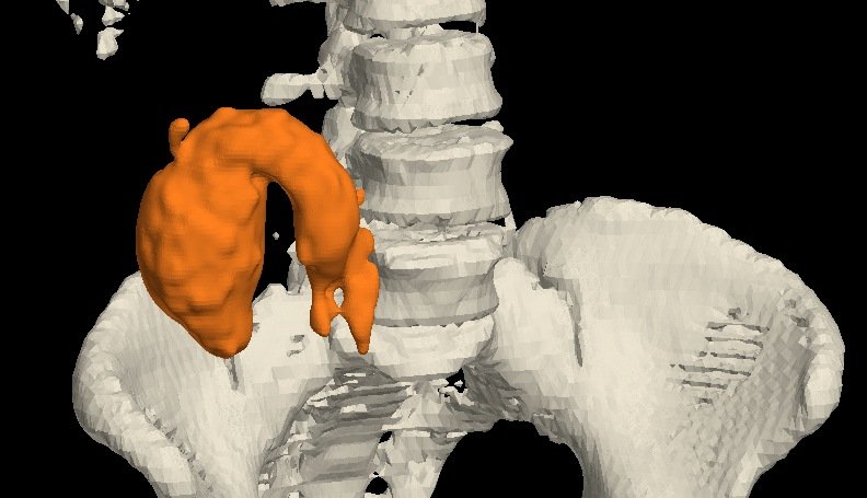
Appendiceal Mucocele: A Fluid-Filled Sausage in the Right Iliac Fossa
Fluid-filled sausage? Think mucocele.
A 50-year-old woman. Contrast CT showed a fluid-filled appendix, 8.9 cm long and 3.8 × 3.9 cm wide, reaching the subhepatic region, with a normal base…
View case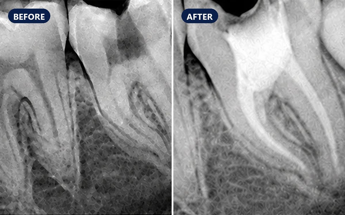
Deep Decay in a Lower Molar, Saved with Root Canal Treatment
Toothache from deep decay? Save the tooth.
A patient had pain from a lower back tooth whose deep decay had reached the nerve (pulp) and caused infection. Root canal treatment removed the infect…
View case
A Toothless Smile Rebuilt with Complete Dentures
No teeth left? Complete dentures give back the smile.
An older man with no teeth left came to the clinic. Before treatment his lips folded inward over the gums; with new upper and lower complete dentures …
View case
Two Canals, Then One, Then Two: A Vertucci Type VI Root Canal
One root, more than one canal? Find and fill every branch.
A lower tooth needed root canal treatment, and its single root held an unusual canal system: two canals leaving the pulp chamber, joining partway down…
View case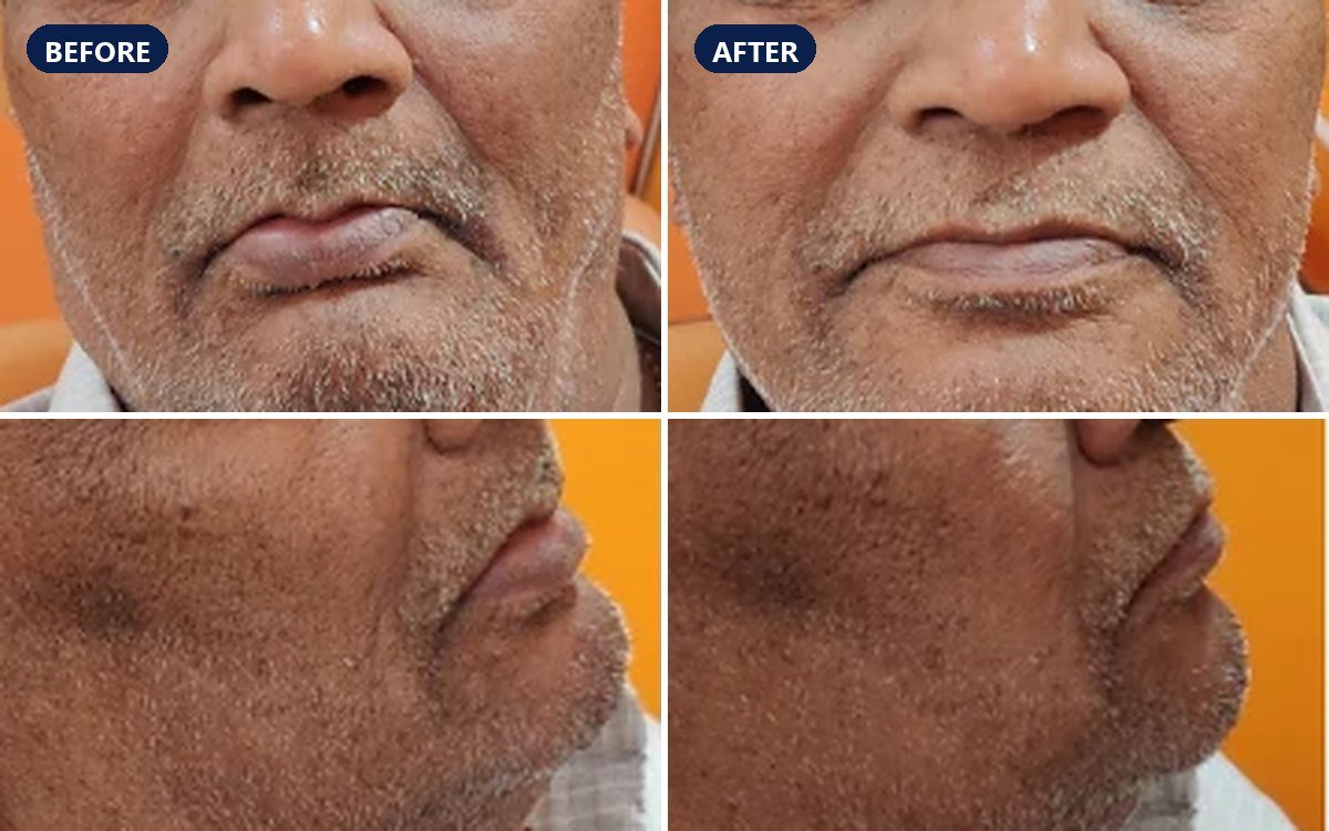
Complete Dentures Restore Lip Support and the Facial Profile
Lips fallen in after losing teeth? Dentures bring back the support.
An older man who had lost all his teeth had also lost the support they give the lips and the lower face. New upper and lower complete dentures put tha…
View case
A Chipped Front Tooth, Rebuilt with Bonded Composite
Chipped front tooth? Bond it back.
An adult patient came to the clinic with a broken edge on an upper front tooth, which left a notch in the smile. The tooth was rebuilt with a direct b…
View case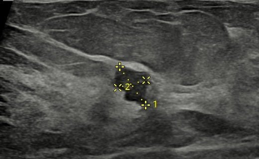
Carcinoma in a Dense Breast: Subtle Distortion, Chased to a Diagnosis
Subtle distortion? Chase it.
A 37-year-old woman with vague left breast pain for a few weeks. Mammography showed heterogeneously dense breasts with mild architectural distortion a…
View case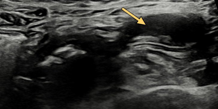
A Twist in the Tail: Meckel's Diverticulum Torsion
Pain and vomiting? Find the blind-ending loop.
A 21-year-old woman with acute abdominal pain and vomiting. Ultrasound showed a dilated pelvic loop with absent flows and an adjacent blind-ending tub…
View case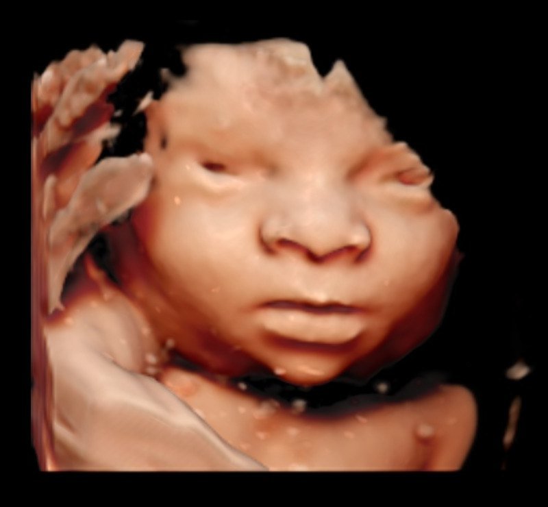
Meet Our HERA W10: A Dedicated Fetal Ultrasound Machine
Our pregnancy scans now run on a Samsung HERA W10, a premium machine dedicated to fetal medicine, with 5D tools, STIC 4D fetal echo, MV-Flow Doppler a…
Read article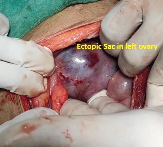
Ovarian Ectopic Pregnancy on Ultrasound
An ectopic in the ovary itself: rare, under 3% of ectopics.
Ultrasound showed a large 6 cm gestational sac in the left adnexa, close to the left ovary, with a fetal pole of 7 weeks 5 days and no cardiac activit…
View case
Torsion of a Meckel's Diverticulum with Gangrene
A dilated pelvic loop and a twisted mesentery: think Meckel's.
Ultrasound showed a focal dilated loop in the pelvis with calcific densities, a twisted mesentery and vessels, and dilated small-bowel loops around it…
View case
Ovary in the Inguinal Canal: Canal of Nuck Hernia
Inguinal swelling in a young woman? The ovary may be inside.
A 20-year-old woman with left inguinal pain and swelling. Ultrasound showed a non-reducible left inguinal hernia containing the left ovary, through a …
View case
A Lump in One Breast, Calcifications in the Other: Why Mammography Matters
A lump in the left breast, and suspicious calcifications in the right.
An 85-year-old woman with a left breast lump. Bilateral mammosonography showed a spiculated malignant-looking lesion with nipple retraction in the lef…
View case
Small VSD on Fetal Echocardiography
A small hole, caught before birth: to-and-fro flow across the septum.
Fetal echocardiography at 25 weeks 4 days showed a small ventricular septal defect, with to-and-fro flow through the defect on colour Doppler.
View case
Endometrial Polyp on 3D Ultrasound: Adding Colours
One polyp, six views: 3D ultrasound of the uterine cavity.
A rounded endometrial polyp seen in the coronal plane of a 3D ultrasound of the uterus, then rendered in different colour and light modes so that the …
View case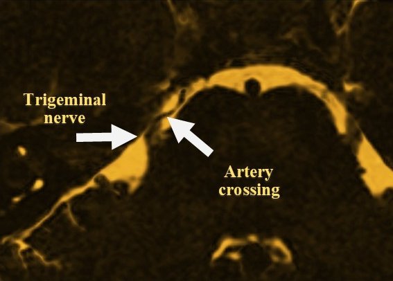
Trigeminal Neuralgia: An Artery Crossing the Nerve on CISS MRI
Electric bursts of facial pain? Look for an artery on the nerve.
Sudden, severe paroxysms of right-sided facial pain lasting seconds to minutes. A high-resolution CISS MRI sequence showed an artery crossing over the…
View case
Non-Contrast MR Renal Angiography
The renal arteries on MRI, with no contrast at all.
A non-contrast MR angiogram shows the abdominal aorta and both renal arteries without any contrast injection: a good way to assess renal artery stenos…
View case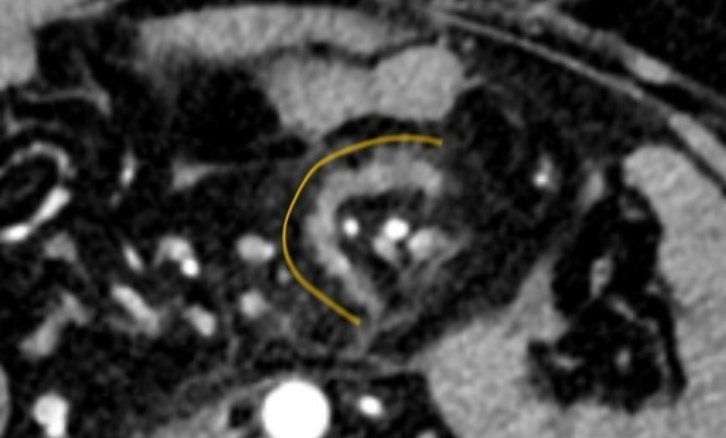
A Tear in the Fan: Traumatic Mesenteric Tear
Blood in the belly, organs intact? Look at the fan.
A 34-year-old man after blunt abdominal injury. Contrast CT showed haemoperitoneum, a C-shaped mesenteric density and a partial SMV thrombus. Laparosc…
View case
Pouches in a Spin: Jejunal Diverticula and Volvulus
No malrotation, yet a whirl? Hunt for the lead point.
A 75-year-old man with pain abdomen and vomiting. Doppler showed a whirlpool of mesenteric vessels; CECT confirmed small-bowel obstruction with an ant…
View case
Uterine & Bowel Perforation after a Failed IUCD Removal
Threads not seen? Scan first.
IUCD threads were not seen and a blind instrument search failed. Next day, ultrasound showed a fundal defect, free fluid and peritoneal thickening. At…
View case
Ileal Diverticulum with a Band: Small-Bowel Obstruction
Small-bowel obstruction with an inflamed pouch? Think diverticulum.
A 58-year-old man with abdominal pain and distension for 2 days. CT showed a diverticulum from a proximal ileal loop with surrounding inflammatory cha…
View case
Hypertrophic Pyloric Stenosis in a 6-Week-Old
Vomiting after every feed? Measure the pylorus.
A 1.5-month-old baby had persistent vomiting after each feed and was not gaining weight. Ultrasound showed the typical pyloric wall thickening of hype…
View case
Jogger's Knee Pain: A Stress Fracture, Not an Osteoid Osteoma
A dense spot on the X-ray after starting to jog? Get the MRI.
A 30-year-old woman had knee pain and difficulty walking for a week after she started jogging. The X-ray raised the question of an osteoid osteoma in …
View case
Peptic Perforation Seen on Ultrasound
Severe epigastric pain? Ultrasound may show the hole.
A 50-year-old man with severe acute epigastric pain. Ultrasound showed echogenic free fluid and a 7 mm defect in the anterior wall of the antropyloric…
View case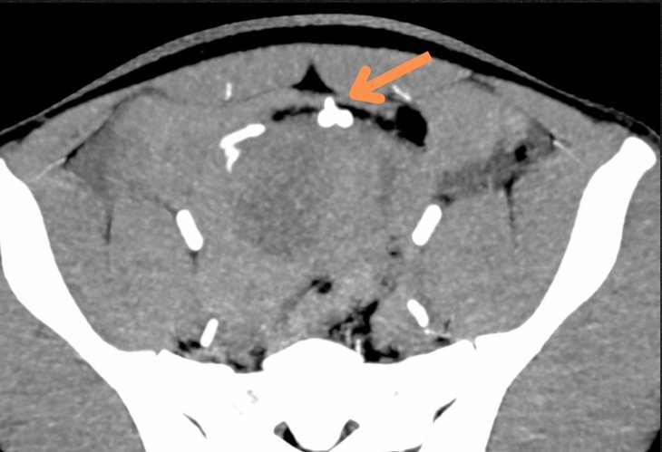
Active Mesenteric Bleed on CT Angiography
Blunt trauma with hypotension? CT angiography finds the bleeder.
A young patient with blunt abdominal trauma and hypotension. CT angiography showed an arterial leak in the pelvis from a mesenteric branch, with activ…
View case
A Large Left Parietal Arteriovenous Malformation
Seizures with arm weakness? Look for the nidus.
A 50-year-old man with seizures and right upper-limb weakness. MRI brain showed a large arteriovenous malformation in the left parietal lobe: a diffus…
View case
The Boomerang Sign: A Splenial Lesion in Hypoglycaemia
Bright splenium after hypoglycaemia? Usually reversible.
A 70-year-old woman suddenly lost consciousness, and lab reports showed hypoglycaemia. MRI brain, done to rule out cerebral injury, showed increased s…
View case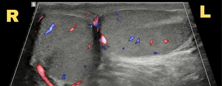
Bell Clapper Deformity: A Testis Lying Sideways
Testicular pain that eased on its own? Check how the testis lies.
Sudden severe pain in the left testis that eased after a few hours. Scrotal ultrasound showed the left testis lying transversely in the sac, the right…
View case
Thyroglossal Fistula: Tracing the Tract on CT Sinogram and MRI
A neck sinus that keeps discharging? Trace the whole tract.
A long-standing sinus in the midline of the neck with occasional pus discharge. A CT sinogram traced the fistula up to the foramen caecum at the base …
View case
Dental Case 1: Rehabilitation in Ectodermal Dysplasia
A female patient with ectodermal dysplasia treated with a Fixed Partial Denture — see the before and after.
Read article
Case 1: Ruptured Appendicitis on X-Ray and Ultrasound
An interesting case where ultrasound and X-ray together confirmed acute appendicitis with rupture — later confirmed at surgery.
View caseNo cases match your search.
Need a scan or an appointment?
We’re open Mon – Sat: 9:00 AM – 7:00 PM | Sun: By Appointment Only | 2D Echo: Thu 5–6 PM | MRI / CT / XRAY / OPG / Pathology: 24×7 available — walk in or call ahead.