
Abdomen & kidneys
Belly pain, stones, lumps and routine check-ups.
- Liver and gallbladder
- Kidneys and bladder
- Aorta
The first Samsung RS85 PRESTIGE in Gujarat: liver stiffness and fat measurement, AI help for breast and thyroid lumps, tiny-vessel Doppler and advanced 3D/4D imaging.
Ours is the first Samsung RS85 PRESTIGE in Gujarat, and the first high-end ultrasound in Panchmahal and Dahod.
Reviewed by Dr Abhishek Agarwal, Consultant Radiologist & Director ·
Ultrasound is also called sonography or USG. At Gujarat Diagnostic Care Plus, sonography is done on the Samsung RS85 PRESTIGE and reported by a radiologist. Pregnancy scans are done on a separate machine, the Samsung HERA W10 (see pregnancy scans).
Every ultrasound uses sound waves, but what you can see depends on the machine. The Samsung RS85 PRESTIGE is Samsung's premium ultrasound system. It gives a sharper picture, and special tools turn what used to be an opinion by eye into a measurement. Your scan is done and reported by an experienced radiologist.
Liver stiffness. A scarred liver (fibrosis) is stiffer than a healthy one. S-Shearwave Imaging measures that stiffness while we watch the live ultrasound picture, so we can see the area being measured. It is painless, takes 2 to 3 minutes and is easy to repeat for follow-up.
Liver fat. Fatty liver is usually graded by eye as mild, moderate or severe. TAI, TSI and EzHRI give a measured number instead, which helps people with fatty liver disease track change over time.
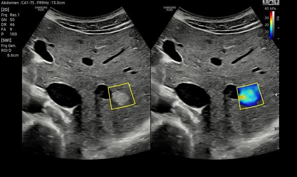
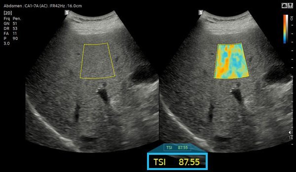
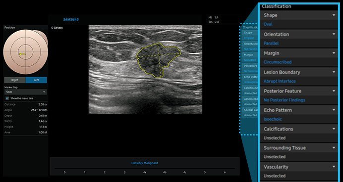
S-Detect is an AI tool that analyses a selected lump and gives a standard report based on recognised breast and thyroid guidelines. E-Breast and E-Thyroid add a check of how stiff the lump is. These tools support the radiologist, who makes the final reading.
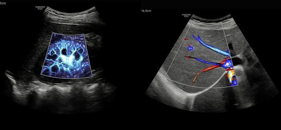
MV-Flow shows slow blood flow in tiny vessels, and LumiFlow gives blood vessels a 3D-like look that is easier to understand. S-Flow picks up flow even in small vessels at the edge of an organ, and Arterial Analysis helps assess the arteries.
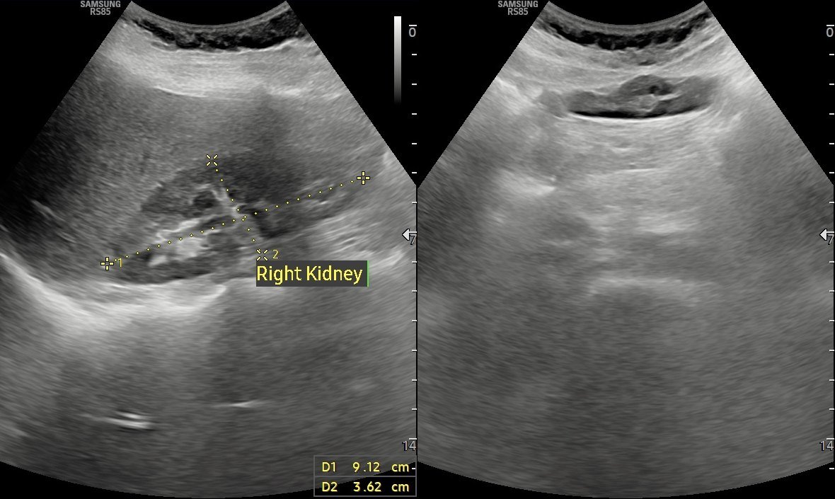
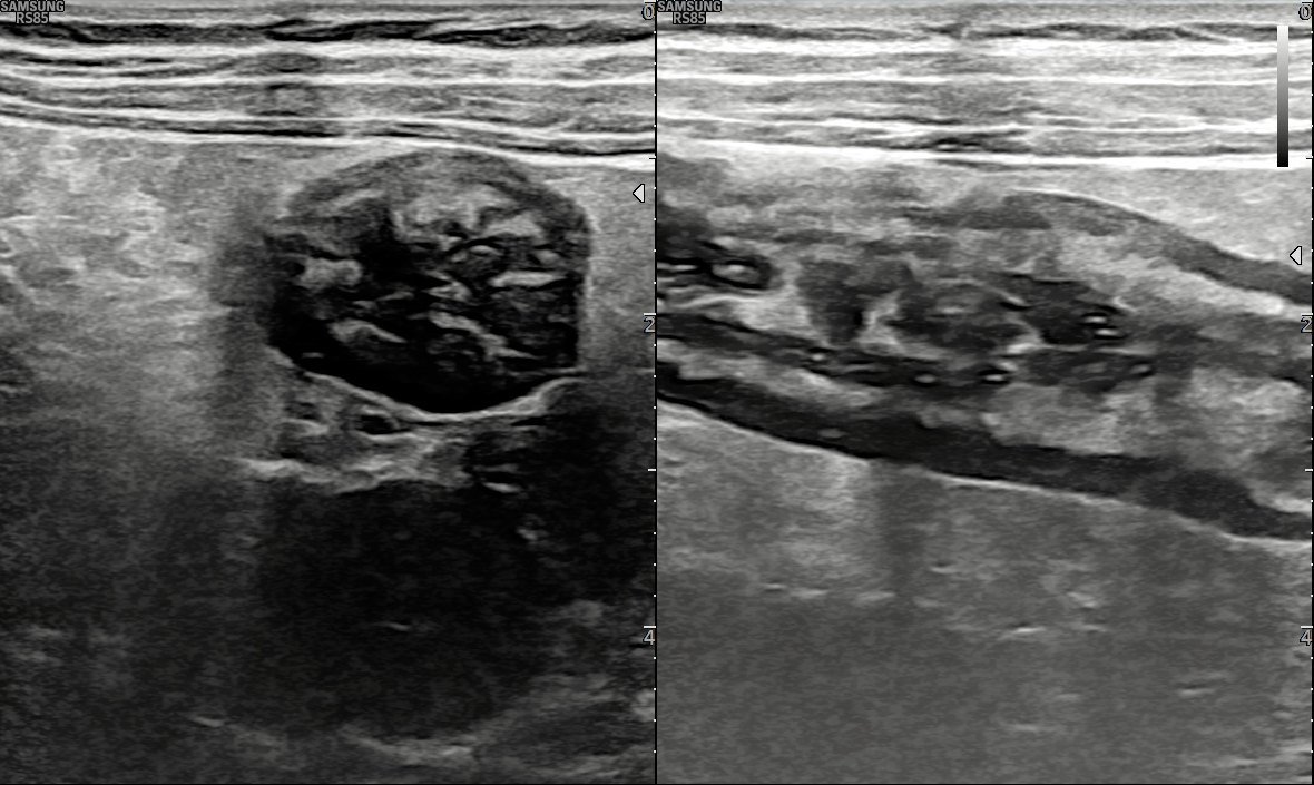
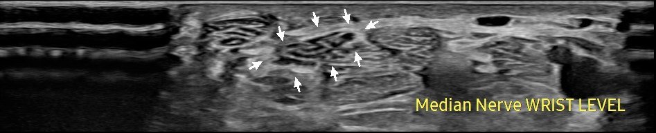
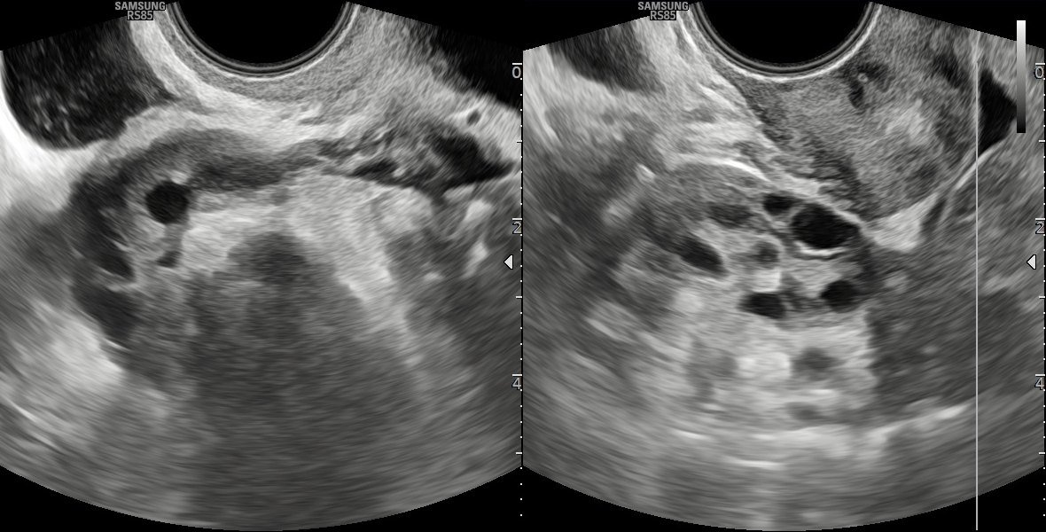
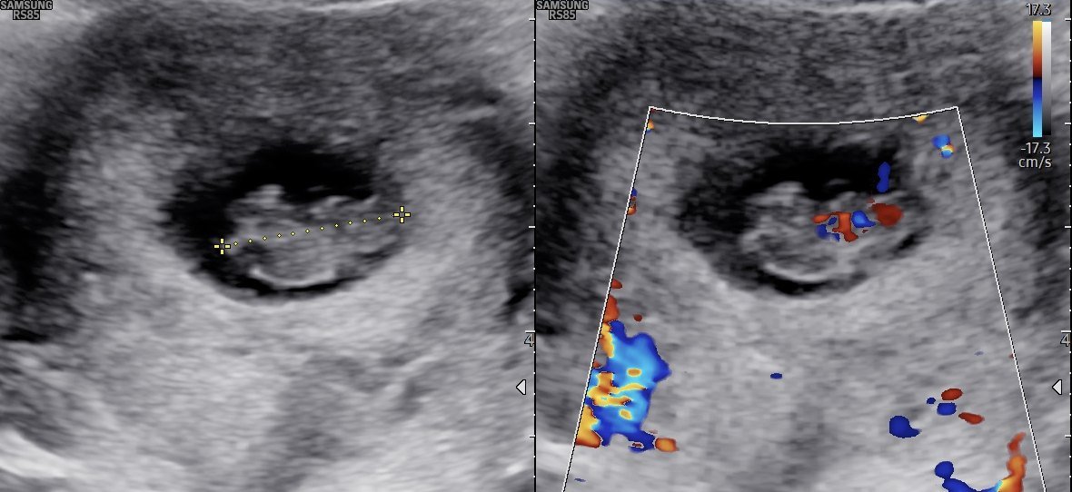
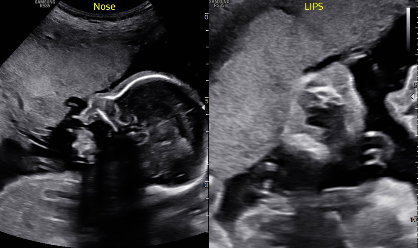
Our pregnancy and fetal scans are done on a dedicated Samsung HERA W10, a premium machine built for obstetrics and fetal medicine, with 5D tools, STIC 4D fetal echo and MV-Flow Doppler. See our Fetal Medicine page.
▸ Fetal 3D/4D Gallery – see the 3D/4D images captured on our high-end machines. Thanks to the babies for giving the poses!
▸ Shearwave Elastography – learn what it is and how it is useful in day-to-day practice.
Sex determination of the fetus is not done at this centre. It is prohibited and punishable under the PCPNDT Act, 1994.
A sharper picture of organs and small structures, and ShadowHDR helps show areas that normally sit behind a shadow.
S-Shearwave measures how stiff the liver is, a sign of scarring, without a needle.
TAI, TSI and EzHRI measure liver fat, so change can be followed over time.
S-Detect analyses breast and thyroid lumps. Our radiologist makes the final reading.
MV-Flow and LumiFlow show slow flow in very small vessels.
Arterial Analysis helps assess the arteries during a Doppler scan.
Not sure which scan you need? Your doctor's request will say, or call us and we will help.
Belly pain, stones, lumps and routine check-ups.
Fatty liver and liver scarring, measured and followed up.
Periods, pain, fertility and lumps in the pelvis.
Lumps and pain, often alongside mammography.
Thyroid nodules and neck lumps.
Pain, swelling and urinary problems.
A close look at the bowel wall with a high-frequency probe.
Tendon, joint and nerve problems.
Doppler checks blood flow and looks for blockages.
Done on our dedicated HERA W10. See the Fetal Medicine page.
Checks blood flow in arteries and veins. Doppler studies are done by appointment.
Scans that need a gentle approach or a specific question answered.
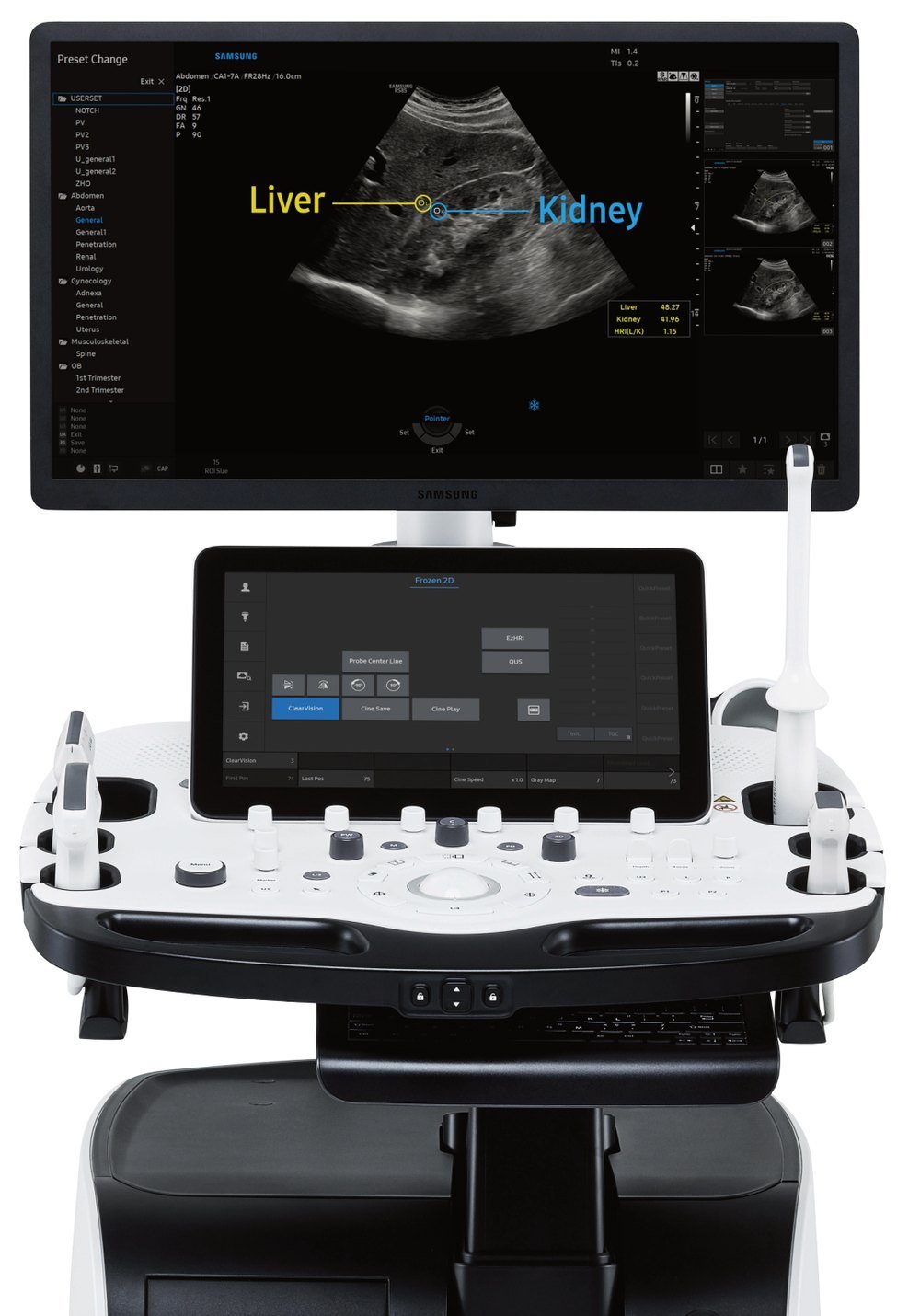
Premium ultrasound system
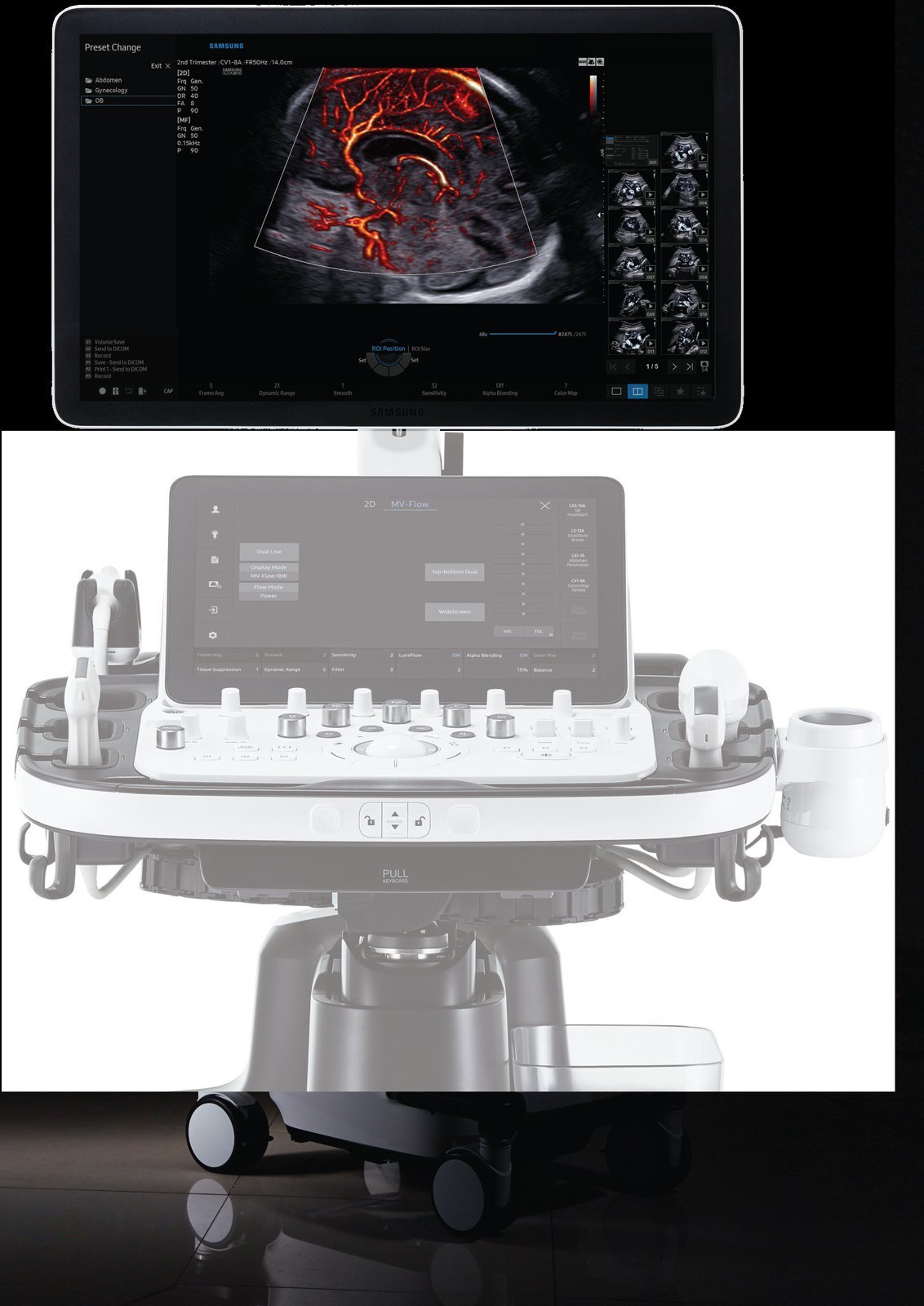
Dedicated pregnancy and fetal ultrasound
Call us with your doctor's request. We will tell you how to prepare.
Our team checks your details and explains the scan.
Gel is put on the skin and a probe moved gently over the area. It is usually painless and generally takes 30 to 45 minutes.
A radiologist reports your scan. Results are usually ready in 24 to 48 hours, and urgent cases are prioritised.
Keep an empty stomach for at least 4 hours and drink water so that the bladder is full.
Prepare as for an abdominal scan. We will confirm when you book.
Fasting for 12 hours is required.
Bring your doctor's request and any earlier scans or reports, so they can be compared.
Your doctor may give you different instructions for your scan; follow those.
Published cases from our own ultrasound work, with patient details removed. Tap an image to enlarge.
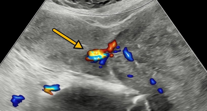
A 38-year-old woman had an 11 cm mass filling the neck of the womb. Colour Doppler traced its stalk back to the lining of the womb, pointing to a fibroid on a stalk. Surgery removed it and confirmed the scan finding, and a 3D model shows the anatomy.
Read the case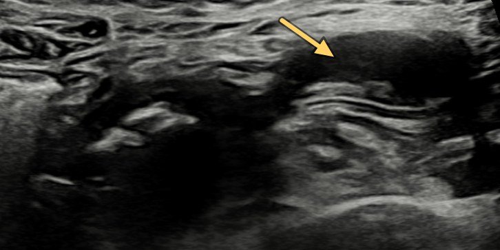
Belly pain and vomiting at 21. Ultrasound with Doppler found a blind-ending loop beside a dilated loop with no blood flow, and a normal appendix. Laparoscopy confirmed a Meckel's diverticulum with a twisted, gangrenous loop.
Read the case
Severe upper belly pain. Ultrasound showed free fluid and a 7 mm defect in the stomach wall, which was repaired at surgery.
Read the case
Ultrasound and X-ray together showed appendicitis with rupture, later confirmed at surgery.
Read the case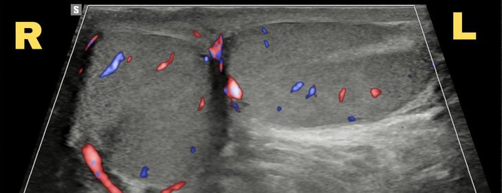
Ultrasound showed a bell clapper deformity, where the testis lies sideways in the sac. It was treated with surgery to fix it in place.
Read the case
Ultrasound showed a 6 cm sac beside the left ovary. Surgery confirmed an ovarian ectopic pregnancy, a rare type.
Read the case
3D ultrasound shows an endometrial polyp and the whole inside of the uterus in one picture.
Read the caseUltrasound uses sound waves to create images of organs and structures inside your body. It's commonly used for pregnancy, abdominal and soft-tissue examinations.
Ultrasound is very safe and does not use any radiation, making it a preferred method for many types of imaging, especially during pregnancy.
No. Sonography is totally harmless to the fetus, even if performed repeatedly.
For abdominal / pelvic ultrasound: keep an empty stomach (fast for at least 4 hours) and drink water so that the bladder is full. For renal Doppler: fasting for 12 hours is required. Our team will confirm specifics when you book.
For certain types of ultrasound — like pelvic or pregnancy scans — a full bladder is necessary to obtain clearer images. Our team will inform you if this applies.
Preparation varies. For abdominal ultrasounds, fasting may be required. We'll inform you of any specific instructions when you schedule the appointment.
An ultrasound generally takes 30 to 45 minutes, depending on the area being examined.
In most cases, yes. Our technicians may show you the images and explain basic information, though the final interpretation is done by a radiologist.
Ultrasound is non-invasive and typically painless, though the gel used may feel cool.
Results are usually available within 24 to 48 hours. We prioritise urgent cases as needed.
It is a painless ultrasound test that measures how stiff the liver is. A scarred liver (fibrosis) is stiffer than a healthy one. It takes about 2 to 3 minutes and needs no needle.
Our RS85 PRESTIGE has tools called TAI, TSI and EzHRI that give a measured number for liver fat, instead of only grading it by eye. The number can be compared at follow-up scans.
Both measure liver stiffness. Ours is done on a full ultrasound machine, so the radiologist watches the live picture and can see the area being measured. The liver and other organs can be checked in the same visit.
Prepare as for an abdominal ultrasound: keep an empty stomach for at least 4 hours. Our team will confirm when you book.
S-Detect analyses a selected lump and gives a standard report based on recognised guidelines. It supports the radiologist, who makes the final reading.
Most scans are done on our Samsung RS85 PRESTIGE. Pregnancy and fetal scans are done on our dedicated Samsung HERA W10.
We’re open Mon – Sat: 9:00 AM – 7:00 PM | Sun: By Appointment Only | 2D Echo: Thu 5–6 PM | MRI / CT / XRAY / OPG / Pathology: 24×7 available — walk in or call ahead.