
Head, face & neck
Head injury, stroke, sinus, eye and ear problems, neck lumps.
- Brain
- Neck
- PNS (sinuses)
- Orbit (eyes)
- HRCT temporal bone (ears)
- Face 3D
The first 32-slice CT in Halol: a Siemens SOMATOM go.Now scanner with low-dose options, available 24×7.
A CT scan uses X-rays and a computer to build detailed pictures of the inside of your body. It is painless, it takes only minutes, and it can give your doctor answers that a plain X-ray cannot.
Reviewed by Dr Abhishek Agarwal, Consultant Radiologist & Director ·
Think of a loaf of bread. A plain X-ray shows the whole loaf from the outside. A CT scan looks at it slice by slice, so your doctor can see each layer clearly and find problems hidden behind other structures.
The scanner can make up to 32 picture slices in each turn around your body. More slices per turn means an area is covered faster, so the scan is shorter and the pictures are less likely to be blurred by breathing or movement.
CT uses X-rays, so every scan carries a small dose of radiation. This chart compares our scanner’s standard dose with the European reference level, the benchmark used to check that a dose is not higher than it should be. A shorter bar means less radiation.
Our scanner (SOMATOM go.Now) European reference level
A low-dose lung scan uses about 84% less radiation than a routine chest CT on this scanner. Your doctor orders a CT when the answer it gives matters more than the small risk, and the aim is always the lowest dose that still gives a clear picture.
Figures are the manufacturer’s default settings for a 75 kg adult (CTDIvol, Siemens dose datasheet). The dose in a real scan varies with body size and the area scanned.
A low-dose lung scan uses about 84% less radiation than a routine chest CT.
Thin 0.7 mm slices, about the thickness of a credit card, show very fine detail.
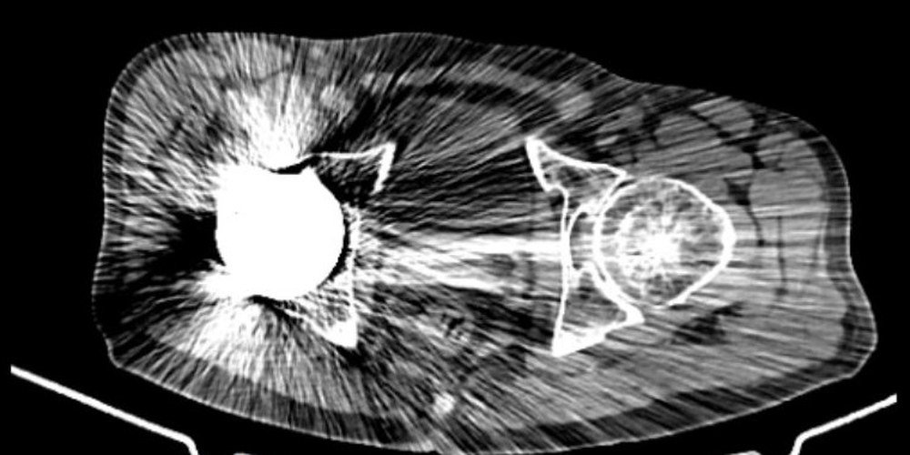
Special software greatly reduces the streaks caused by hip replacements and other metal implants.
The scan usually takes 10 to 15 minutes. The table moves gently through a short, open ring.
Software helps look for small spots in the lungs. Our radiologist makes the final reading.
Bones, joints, blood vessels and teeth can be shown in 3D, which helps doctors plan treatment.
A metal hip can throw bright streaks across a CT picture and hide what lies around it. Slide the handle to compare the same scan before and after our metal reduction software. This is one of our own patients.
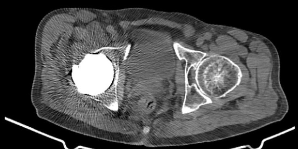
 Before
After
Results vary with the implant and the part of the body.
Not sure which scan you need? Your doctor’s request will say, or call us and we will help.
Head injury, stroke, sinus, eye and ear problems, neck lumps.
Cough, breathlessness, lung infections, lung spots, chest injury.
Belly pain, kidney stones, blockages, injuries, lumps.
Fractures, back and neck pain, joints seen in 3D.
CT angiography shows blocked, narrowed or bleeding vessels.
A 3D view of the jaw and teeth for implant planning.
CT guides a thin needle to take a tiny sample from a lump, usually without a surgical cut.

32-slice CT scanner
Call us with your doctor’s request. CT is available 24×7 for emergencies.
Our team checks your details, explains the scan and tells you if contrast is needed.
Lie still on the table as it moves through the ring. You may be asked to hold your breath for a few seconds. It is painless.
A radiologist reads your scan. Reports are usually ready in 24 to 48 hours, and urgent cases are prioritised.
Some scans need a contrast dye through a vein in the arm. Fasting may be needed; we will tell you when you book. Tell us about any past reaction or allergy.
If you are having contrast and have kidney problems, we may ask for a recent kidney blood test (creatinine).
Tell us if you have diabetes. If you take metformin, ask your doctor whether to pause it around a contrast scan.
Tell us before the scan if you are pregnant or might be. Your doctor may suggest a different test.
Jewellery, hairpins and other metal near the area being scanned will need to come off. Tell us about any implants.
Bring your doctor’s request and any earlier scans or reports, so they can be compared.
Your doctor may give you different instructions for your scan; follow those.
Published cases from our own CT work, with patient details removed. Tap an image to enlarge.
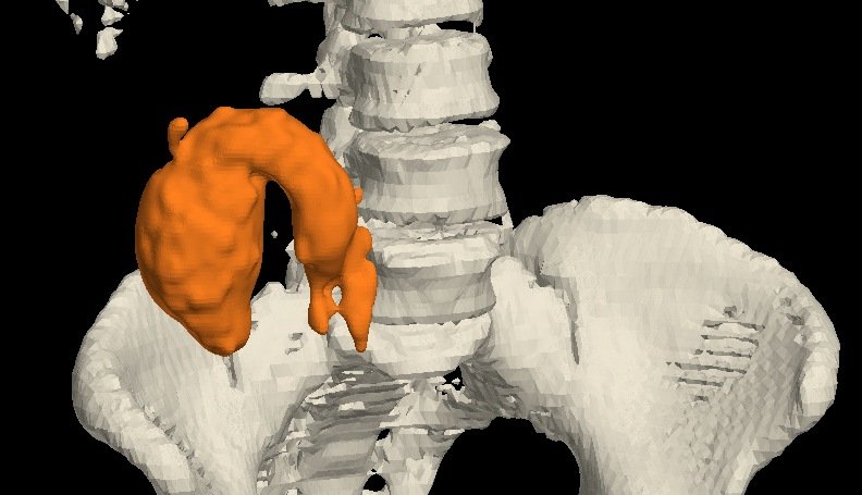
Contrast CT showed an appendix filled with fluid and stretched to 8.9 cm, called a mucocele, and a 3D model was made from the scan. Surgery removed it and confirmed the CT finding.
Read the case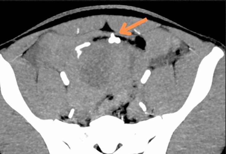
After an injury to the abdomen, CT angiography showed an artery leaking inside the belly. Surgeons could go straight in and tie off the bleeding vessel.
Read the case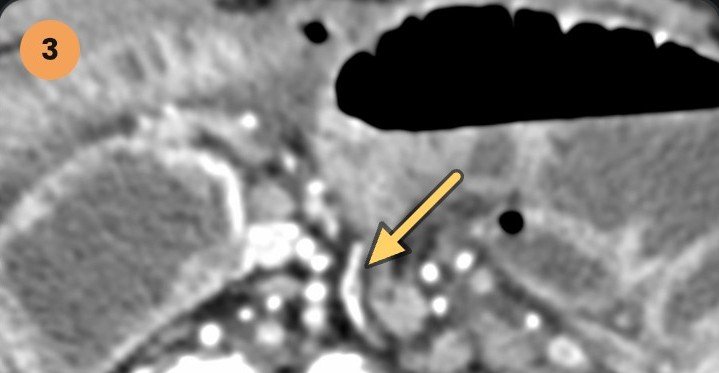
Belly pain and vomiting. CT confirmed a twist in the small bowel causing a blockage. At surgery, small pouches on the bowel wall were found to be the cause.
Read the case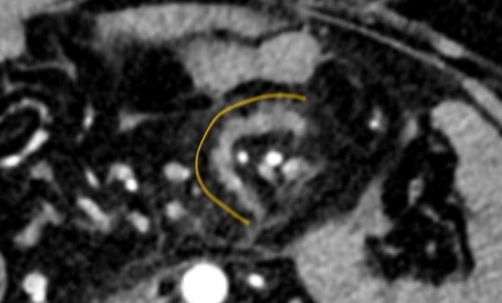
CT showed internal bleeding and damage to the tissue that holds the bowel in place. Surgery found a complete tear and a damaged loop of bowel.
Read the case
Two days of belly pain and swelling. CT showed a small pouch on the bowel with a blockage above it. Surgery confirmed the pouch and a band, and removed them.
Read the case
A CT sinogram followed the tract in 3D, all the way up to the base of the tongue, and MRI added the rest of the picture.
Read the case
An X-ray raised the question of a small bone tumour. MRI found a stress fracture, and CT showed its dense line.
Read the case
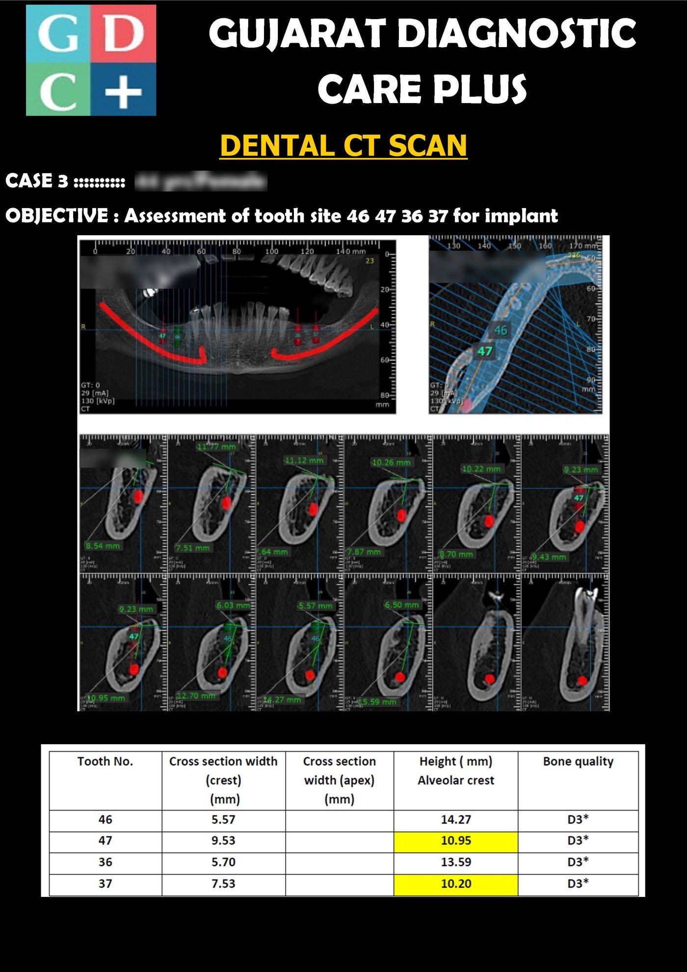
The scan shows the bone available and where the jaw’s nerve runs (traced in red), so the implant can be planned around it.
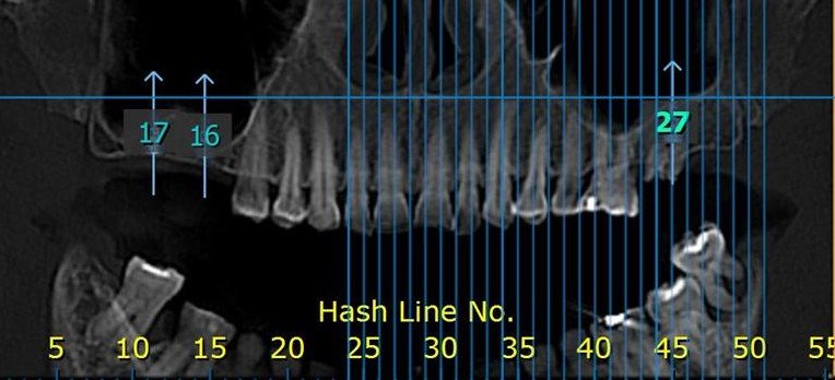
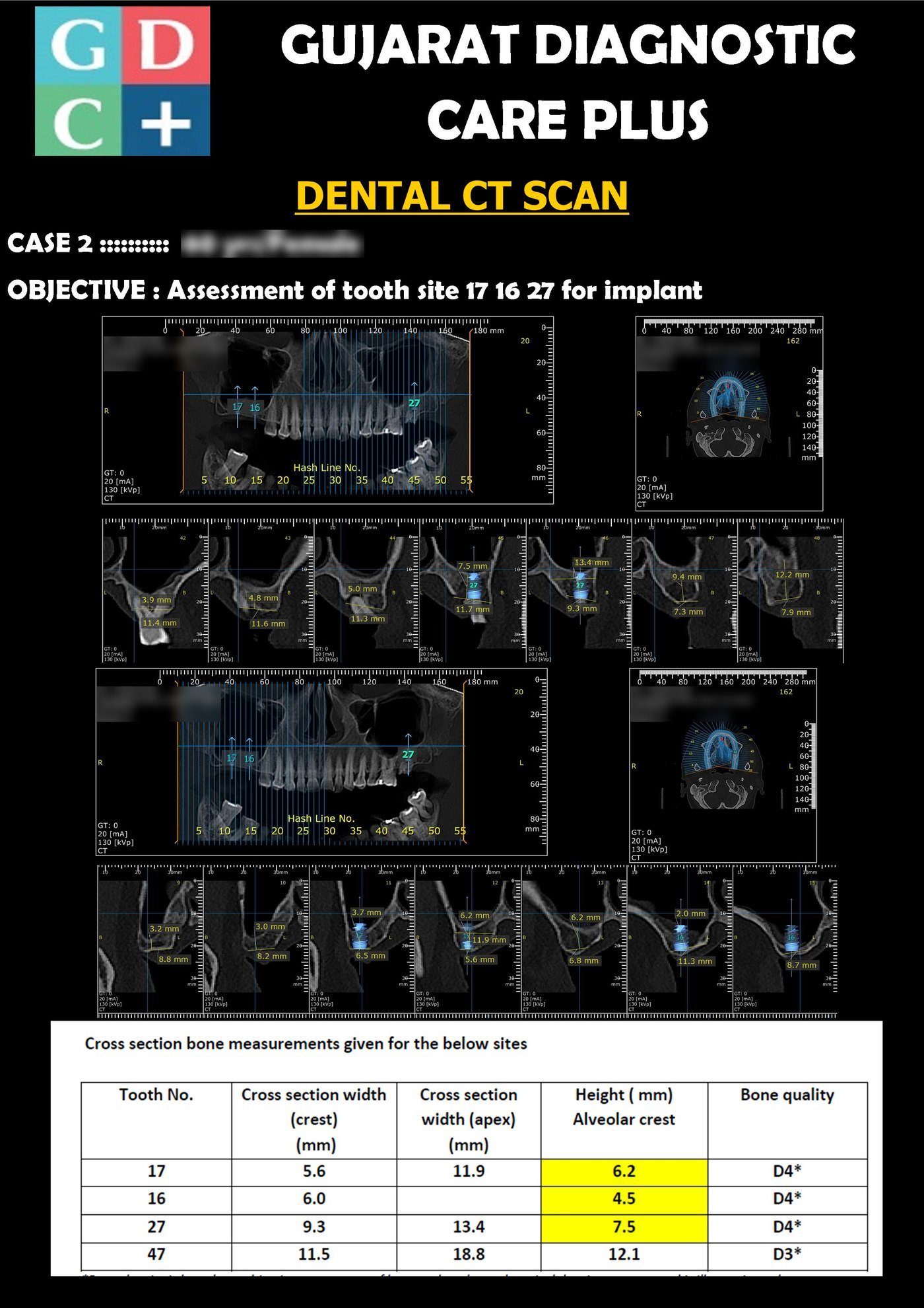
Bone height is measured at each planned site, below the sinus, before the implant is chosen.
A CT scan combines X-ray images from different angles to create detailed cross-sectional images, helping diagnose conditions involving the head, chest, abdomen and bones.
Some CT scans require contrast dye for better visualisation. Our team will inform you if this is needed and explain any specific instructions.
Preparation varies. If contrast dye is used, you may need to fast beforehand. Instructions will be provided at the time of booking.
A CT scan uses more radiation than a regular X-ray, but your doctor orders one only when the information is needed. We keep the dose as low as possible while still getting a clear picture. Our scanner also offers a low-dose lung scan, which uses about 84% less radiation than a routine chest CT.
The scan itself usually takes about 10 to 15 minutes, with the entire appointment lasting about 30 minutes.
For scans without contrast, eating and drinking is allowed. However, fasting may be required if contrast dye is used. Our staff will clarify this when you book.
You'll lie on a table that moves into the scanner. The procedure is painless, and you'll just need to remain still for clear images.
Results are typically ready within 24 to 48 hours, with urgent cases prioritised.
Tell us before the scan if you are pregnant or might be. CT is usually avoided in pregnancy unless it is really needed, and your doctor may suggest ultrasound or MRI instead. In an emergency, your doctor will weigh the benefit against the risk.
Children are more sensitive to radiation, so a CT is done only when the doctor feels it is really needed. Talk to your doctor or to us if you have any worries before the scan.
Yes. Implants such as hip replacements, plates and screws are safe in a CT scanner. Metal can cause streaks in the pictures, and our metal reduction software greatly reduces them. Tell us about any implant before the scan.
Often yes, but tell us first. Contrast leaves the body through the kidneys, so we may ask for a recent kidney blood test (creatinine). If you take metformin, ask your doctor whether to pause it around the scan.
CT uses X-rays and is very quick, which makes it useful for emergencies, injuries, lungs and bones. MRI uses a magnet instead of radiation and shows soft tissues such as the brain, spinal discs and joints in fine detail. Your doctor chooses the test that suits your problem, and we have both under one roof.
A doctor uses CT pictures to guide a thin needle into a lump or abnormal area and take a tiny sample for testing. It is usually done under local anaesthetic, without a surgical cut. You will be told how to prepare, including what to do about blood thinners.

We’re open Mon – Sat: 9:00 AM – 7:00 PM | Sun: By Appointment Only | 2D Echo: Thu 5–6 PM | MRI / CT / XRAY / OPG / Pathology: 24×7 available — walk in or call ahead.