
Baroda Road, Halol Mon–Sat 9 AM–7 PM · MRI, CT, X-ray, OPG and lab 24×7
Waveform Wonders · Interesting case · Neuro

A Large Left Parietal Arteriovenous Malformation

Left parietal AVM → seizures and right arm weakness

Seizures with arm weakness? Look for the nidus.

MRI Dr Abhishek Agarwal · 01 Nov 2026
A Large Left Parietal Arteriovenous Malformation
PatientMale, 50 years, with seizures and right upper-limb weakness
TopicNeuro
ImagingMRI

A 50-year-old man with seizures and right upper-limb weakness. MRI brain showed a large arteriovenous malformation in the left parietal lobe: a diffuse nidus with feeders from the left MCA, PCA and ACA, dilated draining veins, and no aneurysm or haemorrhage.

Case timeline

  1. Presentation

    Seizures and right upper-limb weakness.

  2. MRI brain

    A large arteriovenous malformation in the left parietal lobe on T2W, post-contrast and 3D MRI.

  3. Options

    Microsurgical resection, endovascular occlusion or radiosurgery.

Imaging findings

MRI brain

T2W · post-contrast
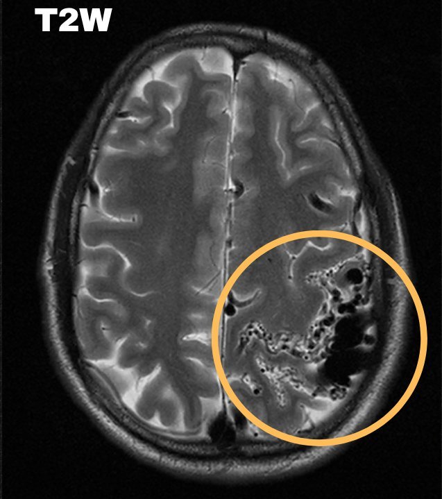
T2W: Tortuous vascular channels in the left parietal lobe (circle).
T2W Tortuous vascular channels in the left parietal lobe (circle).
Post-contrast: The diffuse nidus (circle).
Post-contrast The diffuse nidus (circle).

MRI 3D

Vessel reconstruction
3D MRI: The malformation over the left parietal lobe (arrow).
3D MRI The malformation over the left parietal lobe (arrow).
3D MRI, frontal view: The AVM (arrow) with its feeding arteries.
3D MRI, frontal view The AVM (arrow) with its feeding arteries.
MRI findings

Large left parietal arteriovenous malformation: multiple tortuous arterial and venous channels; a diffuse nidus with early filling of venous channels and scattered calcifications; arterial feeders from the left MCA, PCA and left ACA; multiple dilated, ectatic draining veins. No aneurysm, venous stenosis or haemorrhage in the nidus. Small focal gliotic changes in the fronto-parietal periventricular and deep white matter.

Final diagnosis

Large arteriovenous malformation of the left parietal lobe

Treatment options — microsurgical resection, endovascular occlusion or radiosurgery.
Teaching points

What this case teaches

Take-home message

Seizures with arm weakness? Look for the nidus, and report its feeders, its drainage and any aneurysm or bleed.

How AVMs present

approximate
65%

haemorrhage

20%

seizures, as in this patient

15%

incidental, found in patients with no symptoms

  1. Seizures can be the first sign — about 1 in 5 AVMs present with seizures, as here.

  2. Report the anatomy — nidus, feeding arteries and draining veins, and say whether there is an aneurysm, venous stenosis or haemorrhage (here, none).

  3. Other presentations — haemorrhage is the commonest; headaches and ischaemic events from vascular steal from normal brain also occur.

  4. Treatment — microsurgical resection, endovascular occlusion or radiosurgery.

Need a scan or an appointment?

We’re open Mon – Sat: 9:00 AM – 7:00 PM | Sun: By Appointment Only | 2D Echo: Thu 5–6 PM | MRI / CT / XRAY / OPG / Pathology: 24×7 available — walk in or call ahead.