Baroda Road, Halol Mon–Sat 9 AM–7 PM · MRI, CT, X-ray, OPG and lab 24×7
Waveform Wonders · Interesting case · GI

Active Mesenteric Bleed on CT Angiography

Blunt trauma → mesenteric arterial leak → haemoperitoneum

Blunt trauma with hypotension? CT angiography finds the bleeder.

CTSurgically confirmed Dr Abhishek Agarwal · 01 Nov 2026
Active Mesenteric Bleed on CT Angiography
PatientYoung patient after blunt abdominal trauma, with hypotension
TopicGI
ImagingCT
ProofSurgically confirmed

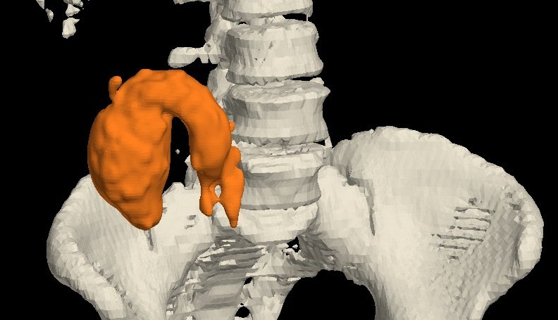
A young patient with blunt abdominal trauma and hypotension. CT angiography showed an arterial leak in the pelvis from a mesenteric branch, with active contrast extravasation that pooled on later phases. Immediate exploration ligated the bleeder and drained 3 litres of haemoperitoneum.

Case timeline

  1. Injury

    Blunt abdominal trauma with hypotension → urgent CT abdomen.

  2. CT angiography

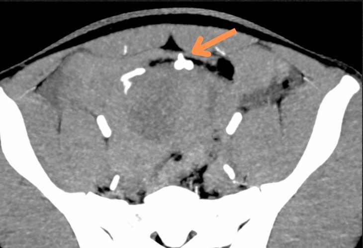
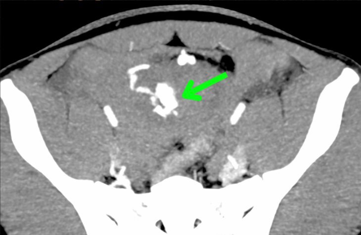
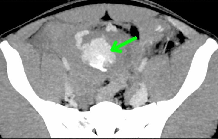
    Arterial leak in the pelvis from a mesenteric branch, with active extravasation of contrast and pooling.

  3. Surgery

    Immediate exploration: the bleeder was ligated and 3 L of haemoperitoneum drained.

  4. Outcome

    The patient's life was saved in time.

Imaging findings

CT angiography

Plain · arterial · venous · delayed
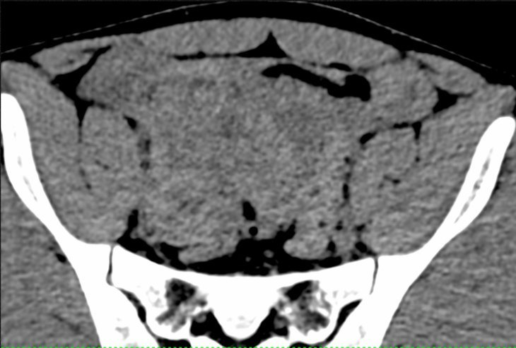
Plain: Before contrast. 1
Plain Before contrast.
Arterial phase: Site of the arterial bleed (orange arrow). 2
Arterial phase Site of the arterial bleed (orange arrow).
Venous phase: Contrast pooling (green arrow). 3
Venous phase Contrast pooling (green arrow).
Delayed phase: Contrast pooling (green arrow). 4
Delayed phase Contrast pooling (green arrow).

The leak shows in the arterial phase (orange arrow), and contrast pools in the venous and delayed phases (green arrows).

CT angiography

Arterial leak in the pelvis from a mesenteric branch, with active extravasation of contrast and pooling.

Final diagnosis

Active arterial bleed from a mesenteric branch after blunt abdominal trauma

Surgery — immediate exploration with ligation of the bleeder; 3 L of haemoperitoneum drained. The patient's life was saved in time.
Teaching points

What this case teaches

Take-home message

Trauma with hypotension? CT angiography finds the bleeder: contrast that leaks in the arterial phase and pools later means active bleeding.

  1. Read the phases together — contrast leaking in the arterial phase and pooling on venous and delayed images means active bleeding.

  2. Find the source — CT angiography plays a vital role in identifying the source of bleeding in trauma patients.

  3. Act early — it guides early intervention: here, immediate exploration and ligation.

Need a scan or an appointment?

We’re open Mon – Sat: 9:00 AM – 7:00 PM | Sun: By Appointment Only | 2D Echo: Thu 5–6 PM | MRI / CT / XRAY / OPG / Pathology: 24×7 available — walk in or call ahead.