Baroda Road, Halol Mon–Sat 9 AM–7 PM · MRI, CT, X-ray, OPG and lab 24×7
Waveform Wonders · Interesting case · GI

A Twist in the Tail: Meckel's Diverticulum Torsion

Meckel's diverticulum → torsion of the adjacent loop → gangrene

Pain and vomiting? Find the blind-ending loop.

UltrasoundDopplerSurgically confirmed Dr Abhishek Agarwal · 12 Nov 2026
A Twist in the Tail: Meckel's Diverticulum Torsion
PatientFemale, 21 years, acute abdominal pain and vomiting
TopicGI
ImagingUltrasound, Doppler
ProofSurgically confirmed

A 21-year-old woman with acute abdominal pain and vomiting. Ultrasound showed a dilated pelvic loop with absent flows and an adjacent blind-ending tubular structure with suspicious twisting of the mesentery, with a normal appendix. Laparoscopy confirmed Meckel's diverticulum with torsion of the adjacent loop and gangrene.

Case timeline

  1. Presentation

    Acute abdominal pain and vomiting → emergency ultrasound of the abdomen.

  2. Ultrasound

    Linear, TVS and Power Doppler: dilated pelvic loop with absent flows and poor wall flow; an adjacent blind-ending tubular structure (? Meckel's diverticulum) with reduced flow and suspicious twisting of the mesentery; the appendix is normal.

  3. Laparoscopy

    Meckel's diverticulum with torsion of an adjacent bowel loop, which was dilated and gangrenous; the gangrenous loop was resected with the Meckel's, with anastomosis.

Imaging & surgical findings

This case includes intra-operative photographs. Tap any image to enlarge it.
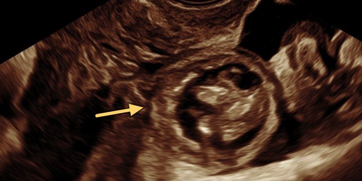
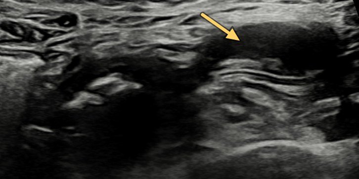
Dilated loop with absent flows: TVS: dilated pelvic bowel loop with absent flows (arrow). 1 Ultrasound
Dilated loop with absent flows TVS: dilated pelvic bowel loop with absent flows (arrow).
Meckel's diverticulum, blind ending: Linear probe: blind-ending Meckel's diverticulum (arrow). 2 Ultrasound
Meckel's diverticulum, blind ending Linear probe: blind-ending Meckel's diverticulum (arrow).
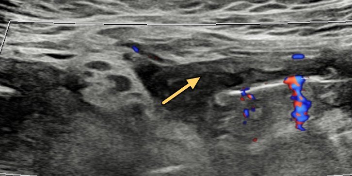
Reduced flow in Meckel's: Power Doppler: reduced flow in the Meckel's diverticulum (arrow). 3 Ultrasound
Reduced flow in Meckel's Power Doppler: reduced flow in the Meckel's diverticulum (arrow).
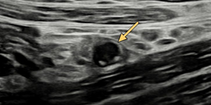
Normal appendix: Linear probe: normal appendix in cross-section (arrow), seen separately. 4 Ultrasound
Normal appendix Linear probe: normal appendix in cross-section (arrow), seen separately.
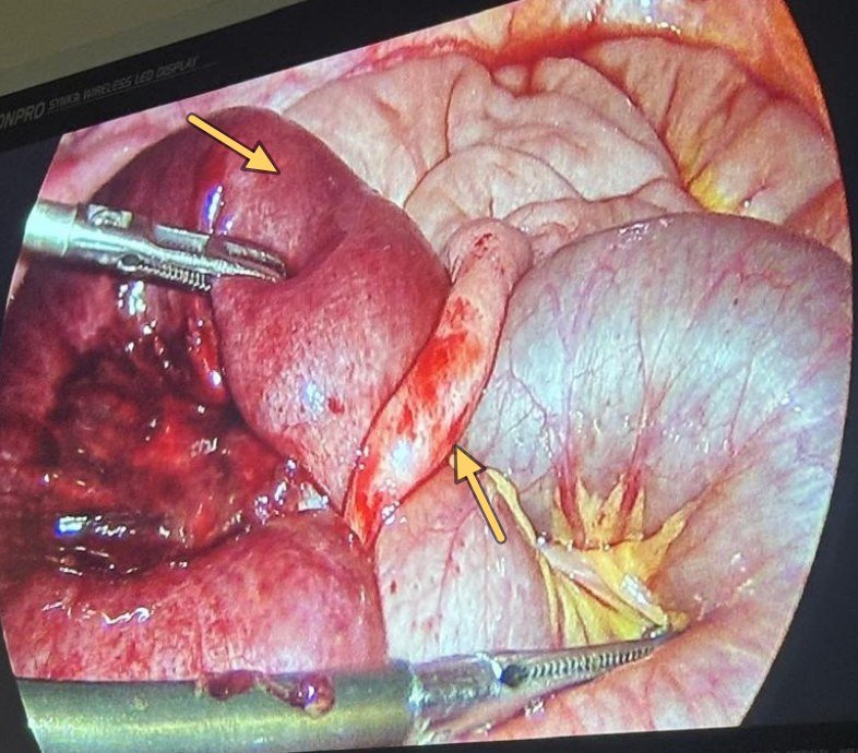
Gangrenous loop and tubular loop: Laparoscopy: dilated gangrenous loop held by a grasper (upper arrow), with the slim tubular loop beside it (lower arrow). 5 Laparoscopy
Gangrenous loop and tubular loop Laparoscopy: dilated gangrenous loop held by a grasper (upper arrow), with the slim tubular loop beside it (lower arrow).
USG impression

Dilated pelvic loop with absent flows, poor wall flow, adjacent blind ending tubular structure with suspicious twisting of mesentery — ? Meckel's with torsion and ischaemic changes.

Final diagnosis

Meckel's diverticulum causing torsion of an adjacent bowel loop, with gangrene

Management — gangrenous loop resected with Meckel's, with anastomosis.

How the twist happens

1

The culprit

A true diverticulum.

2

The pivot

The loop spins around it.

3

Gangrene

Closed loop, no flow.

Teaching points

What this case teaches

Take-home message

Pain and vomiting? Find the blind-ending loop with gut signature, then check its mesentery for a twist.

Rule of 2s

approximate
2%

of people

2 ft

from the ileocaecal valve

2 in

long

2

types of ectopic mucosa

2 yr

age at classic presentation

2:1

male : female

  1. Children bleed, adults obstruct — bleeding from ectopic gastric mucosa; obstruction from volvulus or torsion, intussusception or a Littré hernia.

  2. USG clues — blind-ending, non-compressible tube with gut signature arising from an ileal loop; a normal appendix is seen separately (image 4).

  3. Red flags — a whirl in the mesentery and a thick-walled loop with reduced or absent Doppler flow: think strangulation.

Need a scan or an appointment?

We’re open Mon – Sat: 9:00 AM – 7:00 PM | Sun: By Appointment Only | 2D Echo: Thu 5–6 PM | MRI / CT / XRAY / OPG / Pathology: 24×7 available — walk in or call ahead.