
Case list
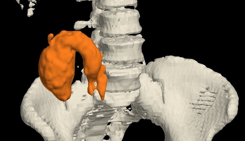
Appendiceal Mucocele: A Fluid-Filled Sausage in the Right Iliac Fossa
Fluid-filled sausage? Think mucocele.
A 50-year-old woman. Contrast CT showed a fluid-filled appendix, 8.9 cm long and 3.8 × 3.9 cm wide, reaching the subhepatic region, with a normal base, no free fluid and no nodes. A 3D model was built from the CT. Surgery removed the grossly distended appendix and confirmed the CT diagnosis.
View case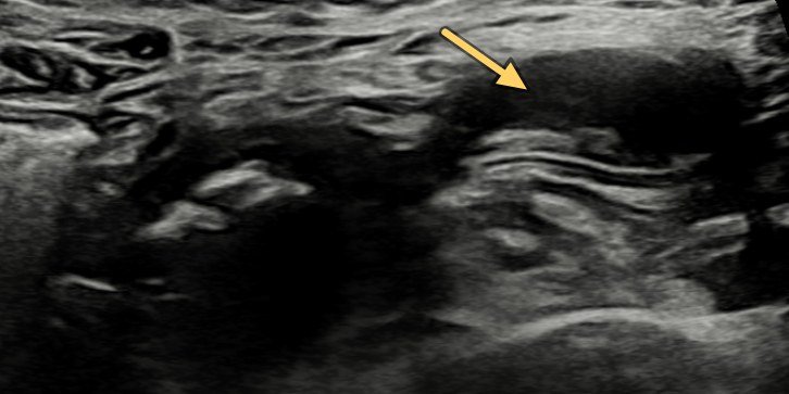
A Twist in the Tail: Meckel's Diverticulum Torsion
Pain and vomiting? Find the blind-ending loop.
A 21-year-old woman with acute abdominal pain and vomiting. Ultrasound showed a dilated pelvic loop with absent flows and an adjacent blind-ending tub…
View case
Torsion of a Meckel's Diverticulum with Gangrene
A dilated pelvic loop and a twisted mesentery: think Meckel's.
Ultrasound showed a focal dilated loop in the pelvis with calcific densities, a twisted mesentery and vessels, and dilated small-bowel loops around it…
View case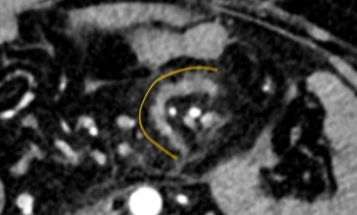
A Tear in the Fan: Traumatic Mesenteric Tear
Blood in the belly, organs intact? Look at the fan.
A 34-year-old man after blunt abdominal injury. Contrast CT showed haemoperitoneum, a C-shaped mesenteric density and a partial SMV thrombus. Laparosc…
View case
Pouches in a Spin: Jejunal Diverticula and Volvulus
No malrotation, yet a whirl? Hunt for the lead point.
A 75-year-old man with pain abdomen and vomiting. Doppler showed a whirlpool of mesenteric vessels; CECT confirmed small-bowel obstruction with an ant…
View case
Ileal Diverticulum with a Band: Small-Bowel Obstruction
Small-bowel obstruction with an inflamed pouch? Think diverticulum.
A 58-year-old man with abdominal pain and distension for 2 days. CT showed a diverticulum from a proximal ileal loop with surrounding inflammatory cha…
View case
Peptic Perforation Seen on Ultrasound
Severe epigastric pain? Ultrasound may show the hole.
A 50-year-old man with severe acute epigastric pain. Ultrasound showed echogenic free fluid and a 7 mm defect in the anterior wall of the antropyloric…
View case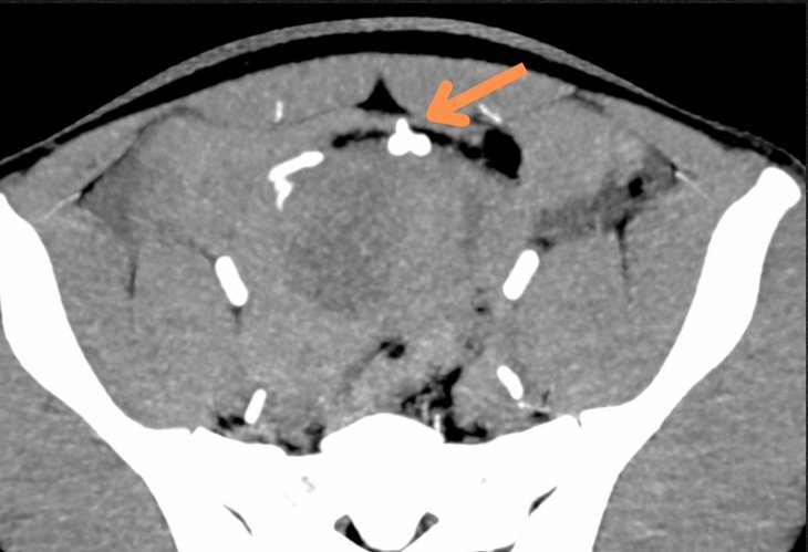
Active Mesenteric Bleed on CT Angiography
Blunt trauma with hypotension? CT angiography finds the bleeder.
A young patient with blunt abdominal trauma and hypotension. CT angiography showed an arterial leak in the pelvis from a mesenteric branch, with activ…
View case
Case 1: Ruptured Appendicitis on X-Ray and Ultrasound
An interesting case where ultrasound and X-ray together confirmed acute appendicitis with rupture — later confirmed at surgery.
View caseNo cases match your search.
Need a scan or an appointment?
We’re open Mon – Sat: 9:00 AM – 7:00 PM | Sun: By Appointment Only | 2D Echo: Thu 5–6 PM | MRI / CT / XRAY / OPG / Pathology: 24×7 available — walk in or call ahead.