
A 37-year-old woman with vague left breast pain for a few weeks. Mammography showed heterogeneously dense breasts with mild architectural distortion and microcalcifications in the left breast. Targeted ultrasound at the same visit found an irregular, stiff 7.8 mm lesion in the left upper outer quadrant, and core biopsy confirmed invasive ductal carcinoma, grade III.
Case timeline
- Presentation
Woman, 37: vague left breast pain for a few weeks; nothing else.
- Mammography
Heterogeneously dense breasts (ACR C); mild architectural distortion with microcalcifications in the upper left breast on the MLO view (D).
- Ultrasound
Same visit, with shear-wave elastography: an irregular, stiff 7.8 mm lesion in the left upper outer quadrant (E, F).
- Core biopsy
USG-guided core biopsy, left breast: invasive ductal carcinoma, NOS — grade III.
Imaging findings
Mammography
4 views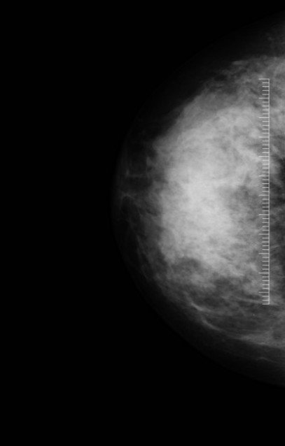 A
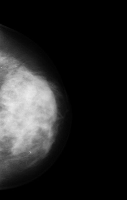 B
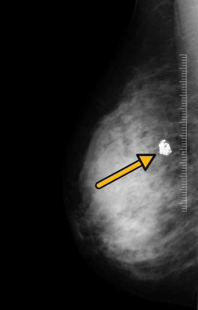 C
 D
Heterogeneously dense breasts (ACR C). Left MLO (D, circle): mild architectural distortion with microcalcifications in the upper breast. Right MLO (C, arrow): coarse ‘popcorn’ calcification — benign.
Look closer
Left MLO, magnified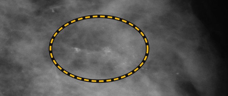 D zoom
Ultrasound
Same visit · shear-wave elastography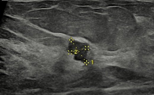 E
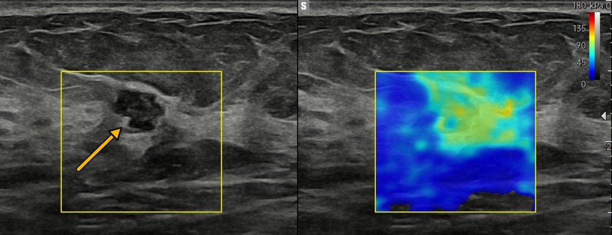 F
Why it looked malignant: irregular shape · not parallel (oblique long axis) · angular margins · stiff on shear-wave (F). Small, yet all four → biopsy.
Invasive ductal carcinoma, NOS — grade III
Histopathology — USG-guided core biopsy, left breast.
Hunt in a dense breast
Architectural distortion
Seen here on the left MLO, upper breast (D).
Fine microcalcifications
Seen here, with the distortion (D zoom).
Focal asymmetry
The third subtle sign. Any one → targeted ultrasound.
