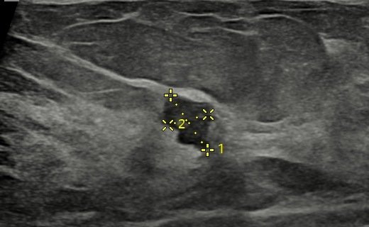
An 85-year-old woman with a left breast lump. Bilateral mammosonography showed a spiculated malignant-looking lesion with nipple retraction in the left breast, and a cluster of calcifications in the right breast, highly suspicious for early DCIS, which could not have been picked up on sonography alone.
Case timeline
- Presentation
Left breast lump → bilateral mammosonography.
- Left breast
Obvious hard lump: spiculated lesion with nipple retraction, a malignant picture on mammography and sonography.
- Right breast
Cluster of calcifications in the outer quadrant, highly suspicious for early DCIS. Not seen on sonography.
Imaging findings
Mammography
Both breasts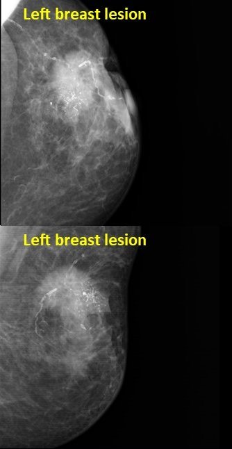

Ultrasound
Left breast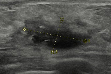
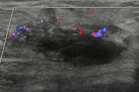
Left: spiculated lesion with nipple retraction, a malignant picture. Right: cluster of calcifications in the outer quadrant, highly suspicious for early DCIS.
Malignant-looking left breast lesion, with right-breast calcifications suspicious for early DCIS
The right-breast calcifications cannot be picked up on sonography alone.