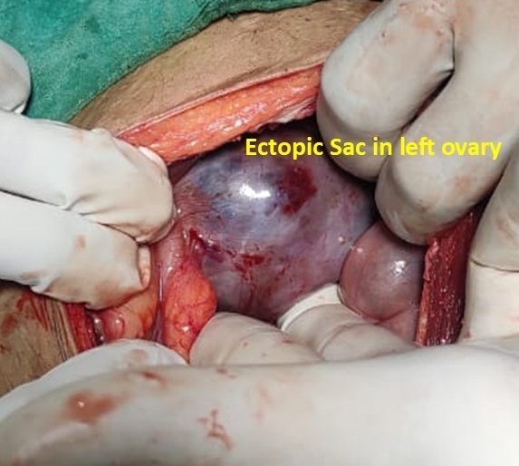
A 20-year-old woman with left inguinal pain and swelling. Ultrasound showed a non-reducible left inguinal hernia containing the left ovary, through a wall defect, with preserved vascularity. Surgery confirmed a canal of Nuck hernia with the ovary in the sac.
Case timeline
- Presentation
Left inguinal pain and swelling → ultrasound.
- Ultrasound
Left inguinal hernia with the ovary as contents, non-reducible, through a wall defect.
- Doppler
Preserved vascularity of the left ovary, which is mildly bulky.
- Surgery
The ovary in the hernia sac.
Imaging & surgical findings
This case includes intra-operative photographs. Tap any image to enlarge it.
Ultrasound
Greyscale · colour Doppler

Surgery

Left inguinal hernia with the ovary as contents, non-reducible. Preserved vascularity of the left ovary, which is mildly bulky.
Left inguinal ovarian hernia (canal of Nuck hernia)
Surgery confirmed the ovary in the hernia sac.