
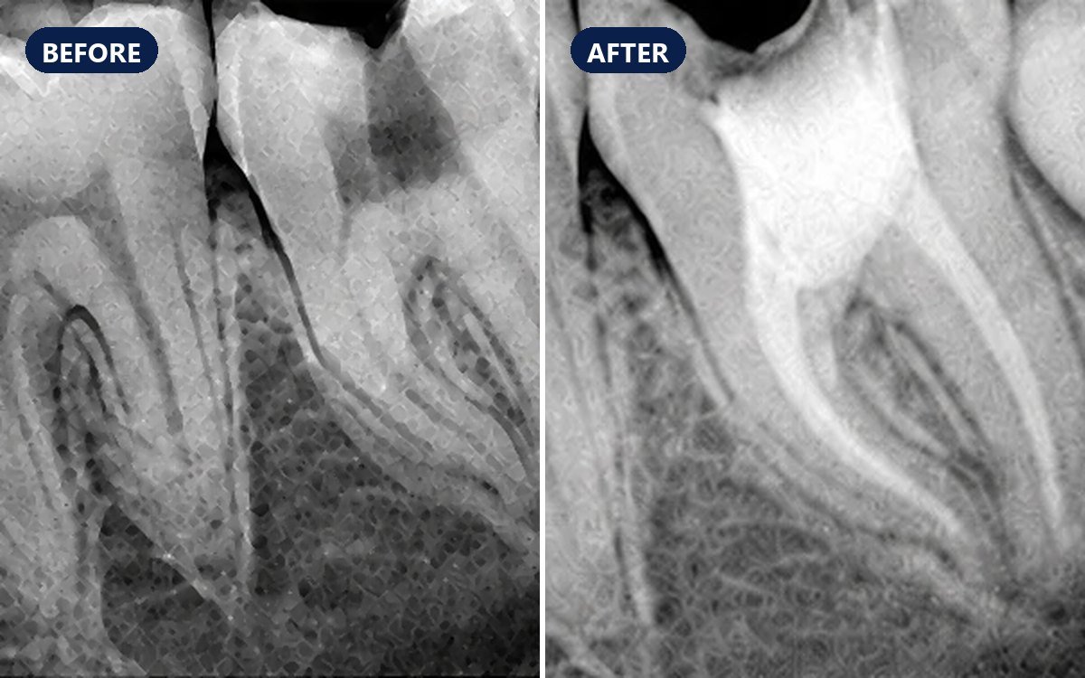
A patient had pain from a lower back tooth whose deep decay had reached the nerve (pulp) and caused infection. Root canal treatment removed the infected pulp, cleaned and filled the canals, and kept the natural tooth. X-rays before and after treatment show the decay and the filled canals.
Case timeline
- Presentation
Pain from a lower back tooth (molar) with deep decay.
- Examination
Intra-oral X-ray (A): a dark area of deep decay in the crown, reaching towards the pulp.
- Treatment
Root canal treatment: the infected pulp removed, the canals cleaned, shaped and disinfected, then filled and sealed.
- Result
X-ray after treatment (B, C): the crown sealed and the canals of both roots filled towards their tips. The tooth was kept and the pain relieved.
Dental X-rays (IOPA)
Before and after
Intra-oral X-rays (IOPA)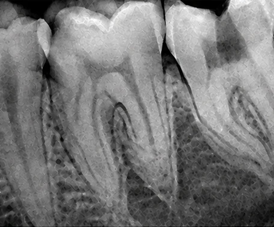
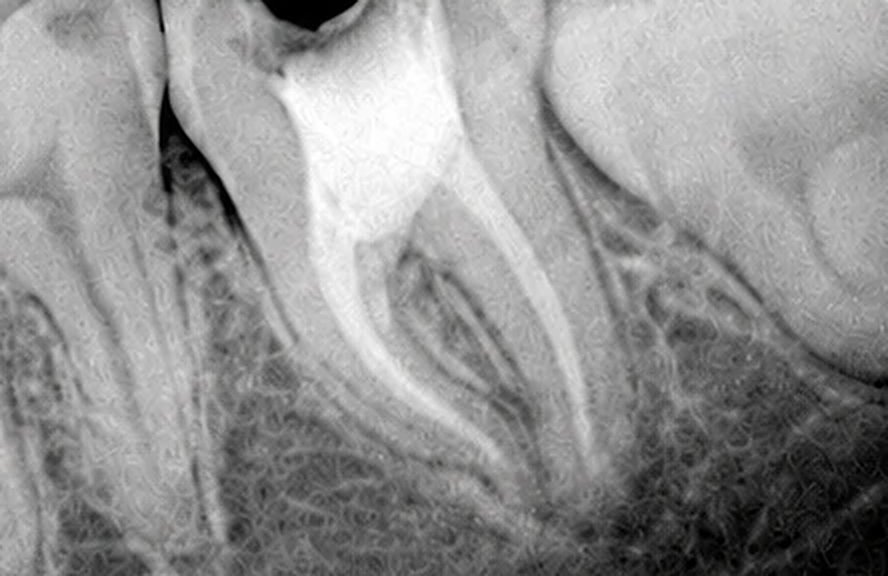
Look closer
The filled canals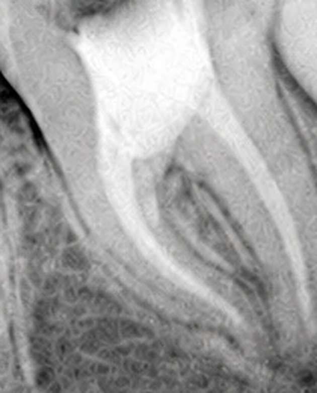 C
Root filling shows white on an X-ray because it blocks X-rays. A good filling runs smoothly, without gaps, to close to the root tip.
Deep dental caries with pulp infection in a lower molar
Treated with root canal (endodontic) treatment and a filling to seal the crown.
How root canal treatment is done
Numb and isolate
The tooth is numbed with local anaesthetic and usually isolated with a thin rubber sheet (rubber dam) to keep it clean and dry.
Clean and shape
An opening in the crown reaches the pulp. The infected pulp is removed, each canal's length is measured, and the canals are shaped with fine files and rinsed with disinfectant.
Fill and seal
The canals are filled with gutta-percha and a sealer, and the crown is sealed with a filling. Back teeth usually get a crown afterwards for strength.
Signs a tooth may need root canal treatment
Pain with hot or cold that lingers after the cause is gone.
Pain on biting, or toothache that wakes you at night.
Swelling of the gum or face, or a pimple-like boil on the gum.
A tooth that has turned dark. Sometimes there are no symptoms and only the X-ray shows the problem.
Why save the natural tooth
A natural tooth keeps its own root and bone, and the neighbouring teeth do not drift into a gap.
It avoids the extra steps of replacing an extracted tooth with a bridge or an implant.
A well-sealed tooth after root canal treatment can last for many years.
After root canal treatment
Mild tenderness for a few days is common and settles with simple painkillers.
Avoid chewing hard food on the tooth until it is fully restored.
A molar that has had root canal treatment is more likely to crack, so a crown is usually advised.
