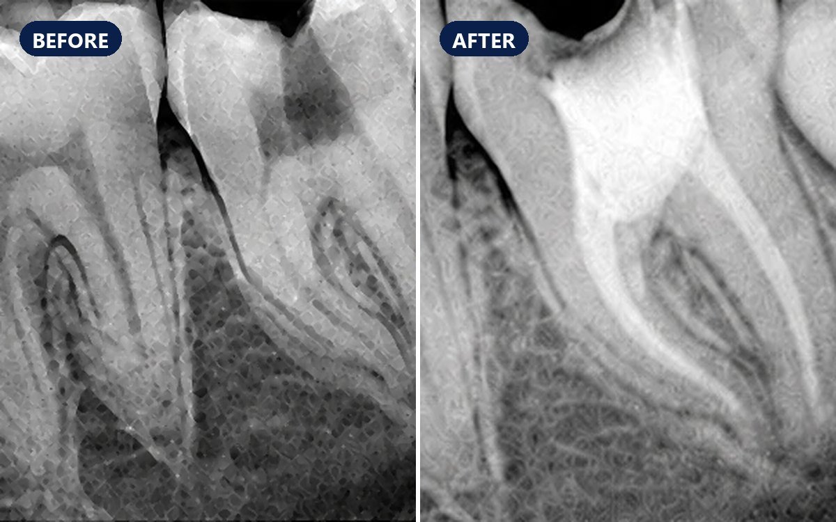
A lower tooth needed root canal treatment, and its single root held an unusual canal system: two canals leaving the pulp chamber, joining partway down the root and dividing again before the tip. This pattern is known as Vertucci Type VI. Each branch was cleaned and filled, and the X-ray after treatment shows the filling tracing the canals.
Case timeline
- Presentation
A lower tooth that needed root canal treatment.
- Anatomy
Inside its single root, the canal system was Vertucci Type VI (2-1-2): two canals that join and then divide again near the root tip.
- Treatment
Root canal treatment, with each canal branch located, cleaned, shaped and filled.
- Result
The X-ray after treatment (A, B) shows the filling running from the pulp chamber down both canals, through the join and on towards the root tip.
Dental X-ray and canal anatomy
After treatment
Intra-oral X-ray (IOPA)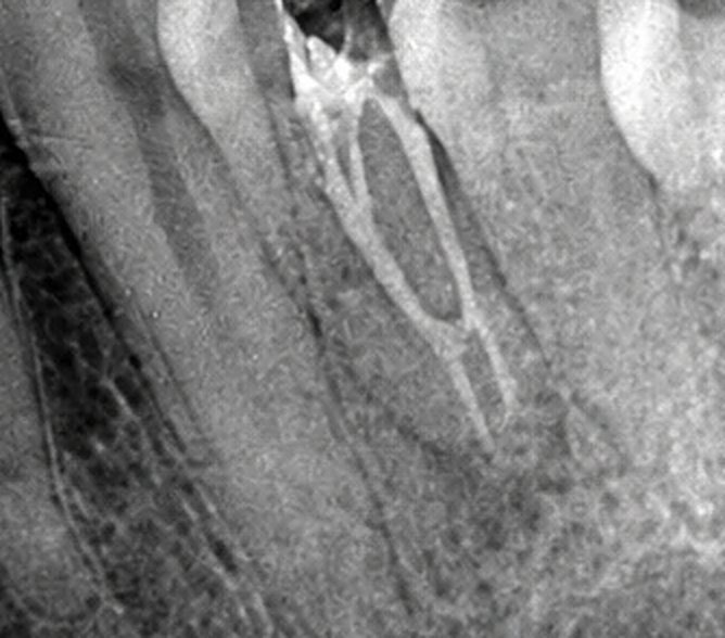 A
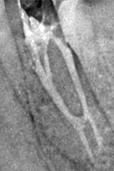 B
The anatomy
Vertucci's classification of root canals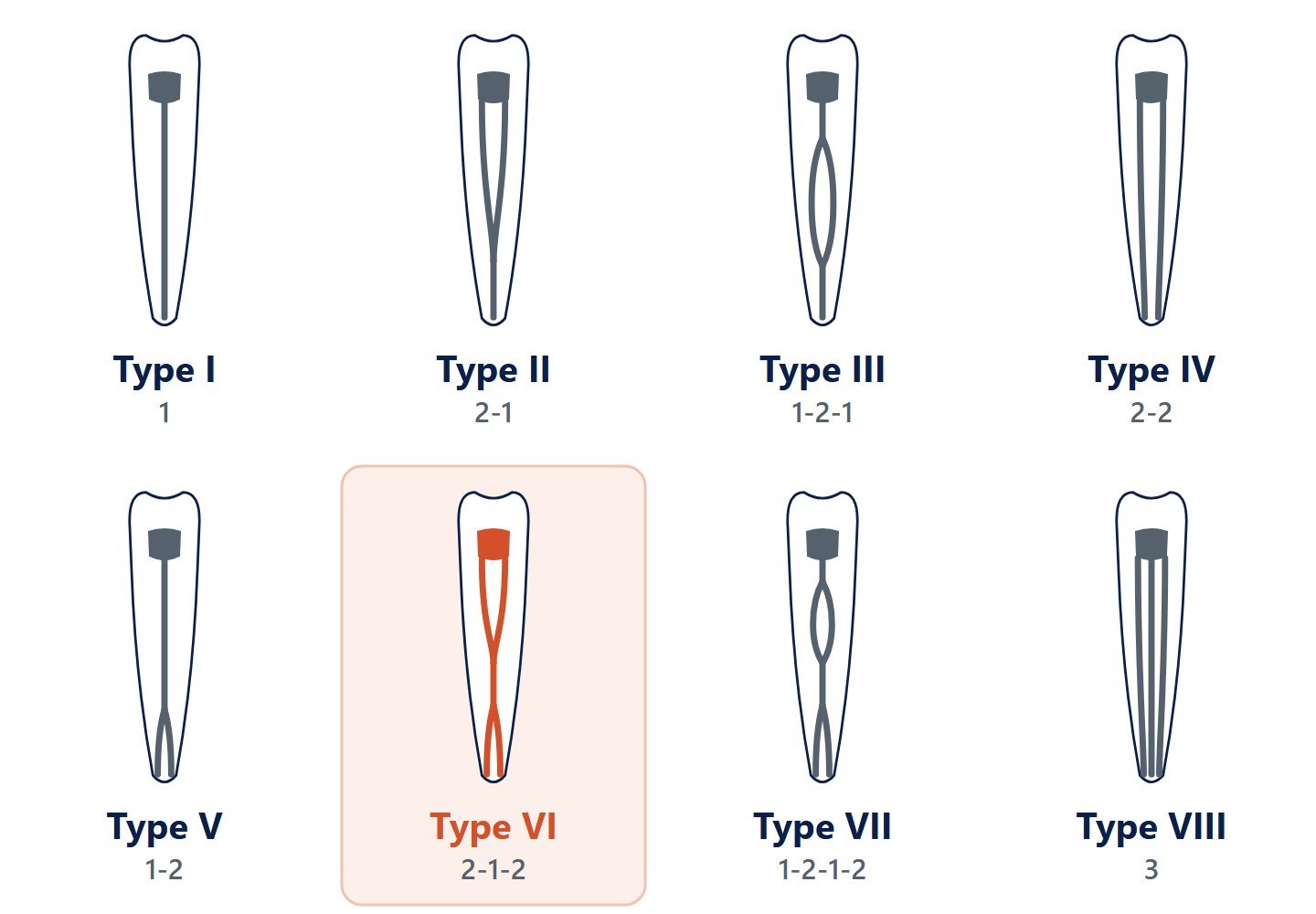 C
Vertucci described eight canal patterns from teeth that were cleared and dye-stained. Most roots hold a single canal; patterns like Type VI are less common and easy to miss on a single X-ray.
Vertucci Type VI (2-1-2) root canal configuration in a lower tooth
Treated with root canal treatment, each canal branch cleaned and filled.
How a complex canal system is treated
Read the X-ray closely
A canal that suddenly narrows or fades partway down the root suggests it divides there. A second X-ray taken from a different angle separates canals that overlap.
Find every branch
The opening into the tooth is shaped to show all the canal entrances, often under magnification. Fine files explore each branch, and an electronic apex locator measures each length.
Clean, shape and fill
Each branch is rinsed with disinfectant, shaped, and filled to its own end, so no part of the canal system is left open to infection.
Vertucci's canal types at a glance
Type I (1): one canal from the pulp chamber to the root tip.
Type II (2-1): two canals join into one before the tip.
Type III (1-2-1): one canal splits in two, then rejoins.
Type IV (2-2): two separate canals all the way.
Type V (1-2): one canal divides into two near the tip.
Type VI (2-1-2): two canals join, then divide again near the tip.
Type VII (1-2-1-2): one canal splits, rejoins and splits again.
Type VIII (3): three separate canals.
Why a missed canal matters
Bacteria left in an untreated branch can keep an infection going after treatment.
A missed canal is a well-known cause of continuing pain or a failed root canal treatment, which is why complex anatomy is looked for before and during treatment.
Where extra canals are common
Some teeth often hold more canals than expected: the front, cheek-side (mesiobuccal) root of upper molars, with its extra MB2 canal, lower front teeth and lower premolars.
In these teeth a second canal is looked for as a routine.