Baroda Road, Halol Mon–Sat 9 AM–7 PM · MRI, CT, X-ray, OPG and lab 24×7
GDC Case Archive · Interesting case · Gynaecology

Endometrial Polyp on 3D Ultrasound: Adding Colours

3D coronal view of the uterine cavity → endometrial polyp

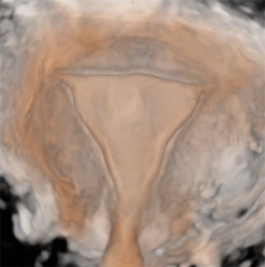
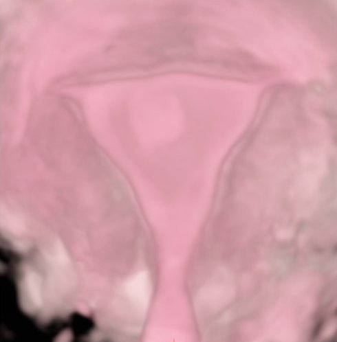
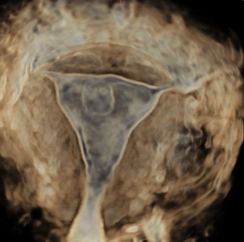
One polyp, six views: 3D ultrasound of the uterine cavity.

3D ultrasound Dr Abhishek Agarwal · 03 Nov 2026
Endometrial Polyp on 3D Ultrasound: Adding Colours
PatientEndometrial polyp seen on 3D ultrasound of the uterus
TopicGynaecology
Imaging3D ultrasound

A rounded endometrial polyp seen in the coronal plane of a 3D ultrasound of the uterus, then rendered in different colour and light modes so that the polyp and the whole uterine cavity are seen in one picture.

Case timeline

  1. 3D ultrasound

    The coronal plane shows a rounded polyp in the endometrial cavity (arrow).

  2. Rendering

    The same 3D volume shown in different colour and light modes.

Imaging findings

Coronal plane
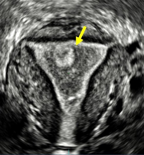
Coronal plane: The polyp (arrow) in the endometrial cavity.
Coronal plane The polyp (arrow) in the endometrial cavity.

3D renderings

Colour and light modes
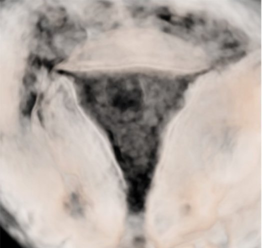
Sepia render: The whole cavity in one view.
Sepia render The whole cavity in one view.
Pink render: Soft surface rendering of the cavity.
Pink render Soft surface rendering of the cavity.
Inverted render: The cavity outlined against the myometrium.
Inverted render The cavity outlined against the myometrium.
Glass render: The polyp seen through the rendered cavity.
Glass render The polyp seen through the rendered cavity.
Light render: The polyp (arrow) in a light-rendered view.
Light render The polyp (arrow) in a light-rendered view.
3D ultrasound

A rounded endometrial polyp within the uterine cavity, seen in the coronal plane.

Final diagnosis

Endometrial polyp on 3D ultrasound
Teaching points

What this case teaches

Take-home message

3D ultrasound turns the uterine cavity into one picture, and a polyp stands out.

  1. Coronal plane — 3D shows the whole cavity, from fundus to cervix, in one view.

  2. Shows the site — the polyp is easy to show to the gynaecologist before hysteroscopy.

  3. Colour renderings — help the patient and the referring doctor see the finding.

Need a scan or an appointment?

We’re open Mon – Sat: 9:00 AM – 7:00 PM | Sun: By Appointment Only | 2D Echo: Thu 5–6 PM | MRI / CT / XRAY / OPG / Pathology: 24×7 available — walk in or call ahead.