Baroda Road, Halol Mon–Sat 9 AM–7 PM · MRI, CT, X-ray, OPG and lab 24×7
Interesting case · Gynaecology

Pedunculated Fibroid in the Cervix: Trace the Stalk

Mass in the cervical canal → stalk traced to the endometrium on Doppler → pedunculated fibroid at surgery

Cervical mass? Trace the stalk.

UltrasoundColour DopplerSurgically confirmed Dr Abhishek Agarwal · 17 Nov 2026
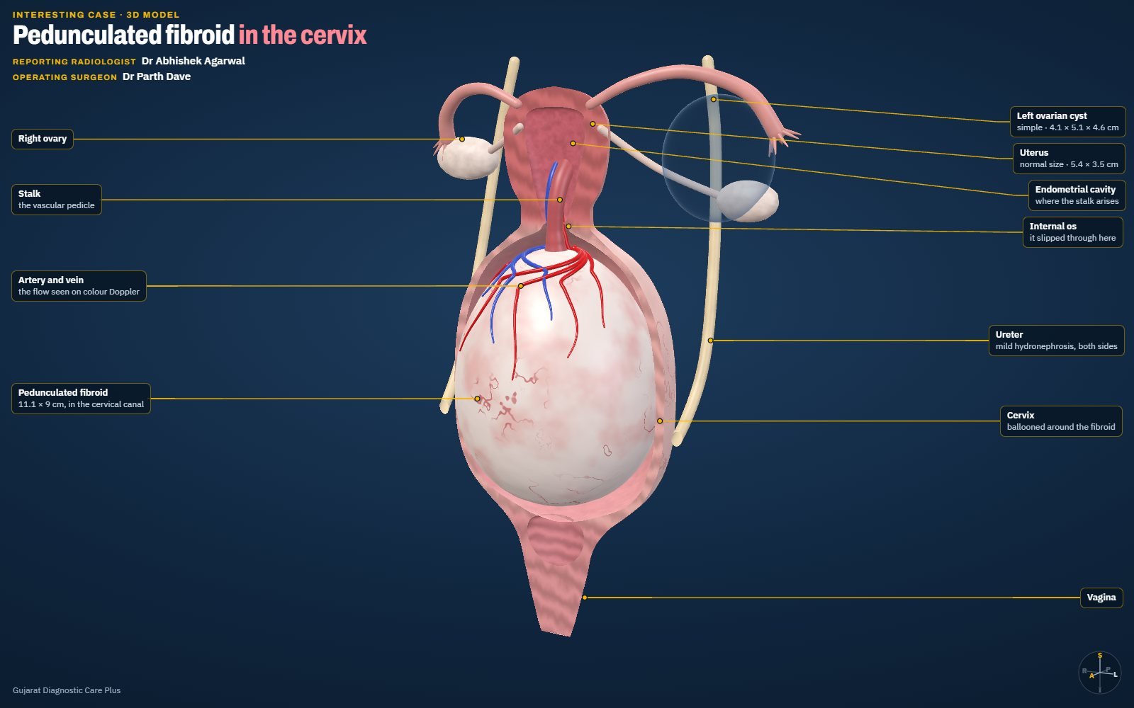
Schematic 3D model: Pedunculated Fibroid in the Cervix: Trace the Stalk
PatientFemale, 38 years
TopicGynaecology
ImagingUltrasound, Colour Doppler
ProofSurgically confirmed

A 38-year-old woman with a Foley catheter in place. Ultrasound showed a 111 × 90 mm mass filling the cervical canal, with mild hydronephrosis on both sides. Colour Doppler traced its stalk to the endometrium, and surgery confirmed a pedunculated fibroid. An interactive 3D model shows how it hangs on its stalk.

Case timeline

  1. Day 0

    Woman, 38, referred for ultrasound; Foley catheter in situ.

  2. USG

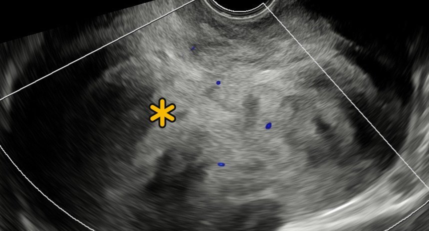
    111 × 90 mm mass in the cervical canal (A, C); mild bilateral hydronephrosis.

  3. Doppler

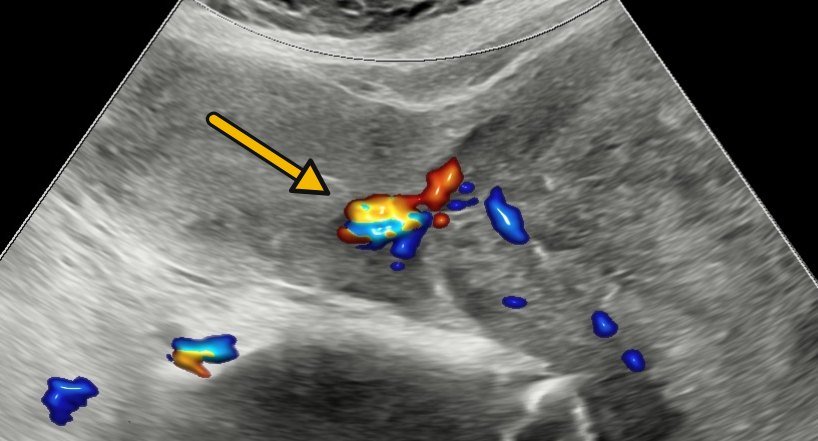
    Vascular pedicle from the endometrium (B) → fibroid or polyp.

  4. Surgery

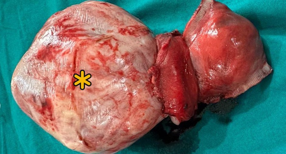
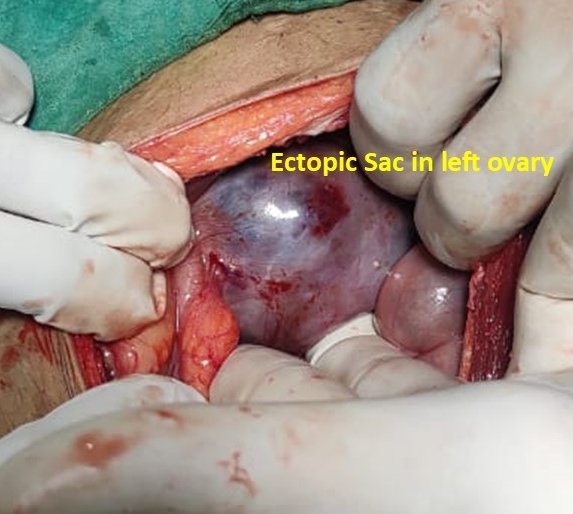
    Pedunculated fibroid (D): the ultrasound diagnosis held.

Imaging & surgical findings

This case includes intra-operative photographs. Tap any image to enlarge it.
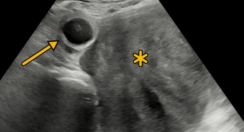
Mass in the cervical canal: Transabdominal ultrasound: heteroechoic mass (*) filling the cervical canal; Foley balloon (arrow). A Ultrasound
Mass in the cervical canal Transabdominal ultrasound: heteroechoic mass (*) filling the cervical canal; Foley balloon (arrow).
Vascular pedicle: Colour Doppler: the stalk (arrow), arising from the endometrium. B Colour Doppler
Vascular pedicle Colour Doppler: the stalk (arrow), arising from the endometrium.
Transvaginal view: Heteroechoic mass (*) with mild internal vascularity. C Ultrasound
Transvaginal view Heteroechoic mass (*) with mild internal vascularity.
Pedunculated fibroid: Post-op specimen (*). Sonography findings confirmed. D Surgery
Pedunculated fibroid Post-op specimen (*). Sonography findings confirmed.
Final diagnosis

Pedunculated fibroid in the cervical canal

Surgery — pedunculated fibroid; the ultrasound diagnosis held.
Interactive 3D model

Explore it in 3D

Schematic teaching model built to the ultrasound report’s measurements: the uterus, the stalk with its artery and vein, the fibroid in the cervical canal, the bladder with the Foley balloon, the ovaries and the ureters. Rotate it, switch views and layers, or slice the fibroid.

Drag to rotate, scroll or pinch to zoom, and switch views and layers. Open full screen

Teaching points

What this case teaches

Take-home message

For a mass in the cervical canal, trace its stalk on colour Doppler: a stalk from the cavity points to a submucous fibroid or polyp, no stalk calls for MRI, and a large mass needs a check for urinary retention and hydronephrosis.

  1. Mass in the cervical canal → colour Doppler: trace its stalk.

  2. Stalk from the cavity → submucous fibroid or polyp. No stalk → MRI.

  3. Large cervical mass → look for urinary retention and hydronephrosis.

Images are de-identified and shared for medical education.

Need a scan or an appointment?

We’re open Mon – Sat: 9:00 AM – 7:00 PM | Sun: By Appointment Only | 2D Echo: Thu 5–6 PM | MRI / CT / XRAY / OPG / Pathology: 24×7 available — walk in or call ahead.