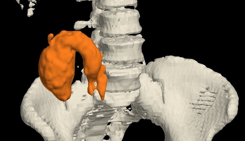
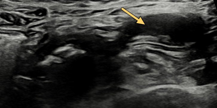
A 75-year-old man with pain abdomen and vomiting. Doppler showed a whirlpool of mesenteric vessels; CECT confirmed small-bowel obstruction with an anticlockwise mid-jejunal twist and no malrotation. At laparotomy, multiple jejunal diverticula were the lead point.
Case timeline
- Day 0
Referred for an abdominal ultrasound for pain abdomen and vomiting. Doppler: whirlpool of mesenteric vessels (1); a diverticulum, seen in retrospect (2); bilateral polycystic kidneys (5, 6).
- Day 2
CECT: acute SBO with multiple jejunal diverticula; abrupt mid-jejunal transition with an anticlockwise mesenteric twist (3, 4). Normal SMA–SMV relation, no mass.
- Day 6
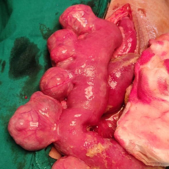
Laparotomy: segmental jejunal volvulus with multiple jejunal diverticula as the lead point.
- Outcome
Diverticula-bearing segment resected with primary anastomosis; uneventful recovery.
Imaging & surgical findings
This case includes intra-operative photographs. Tap any image to enlarge it.
Ultrasound
Day 0 · Doppler + linear probe

CT scan
Day 2 · contrast-enhanced · axial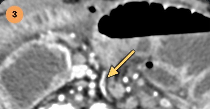
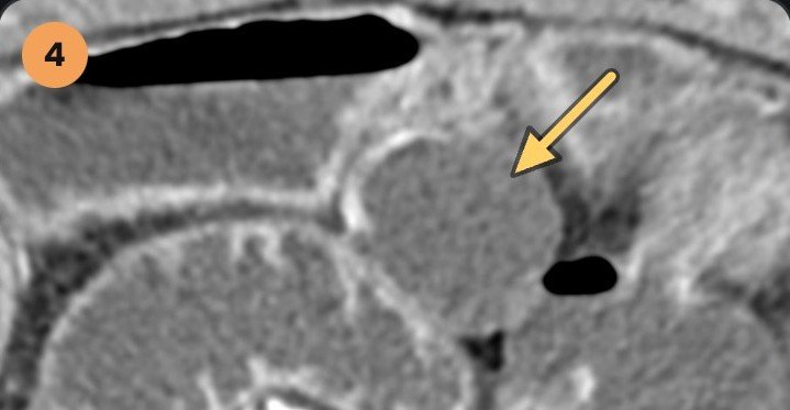
CT cine
Arterial phase · Im 118 → 130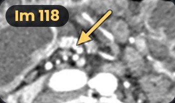
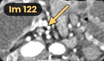
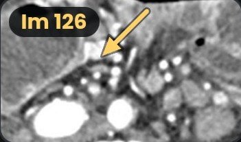
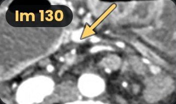
Scrolling down through the root, the vessels wind round the twisted mesentery.
Surgery
Day 6 · laparotomy

Polycystic kidneys
Day 0 · USG · incidental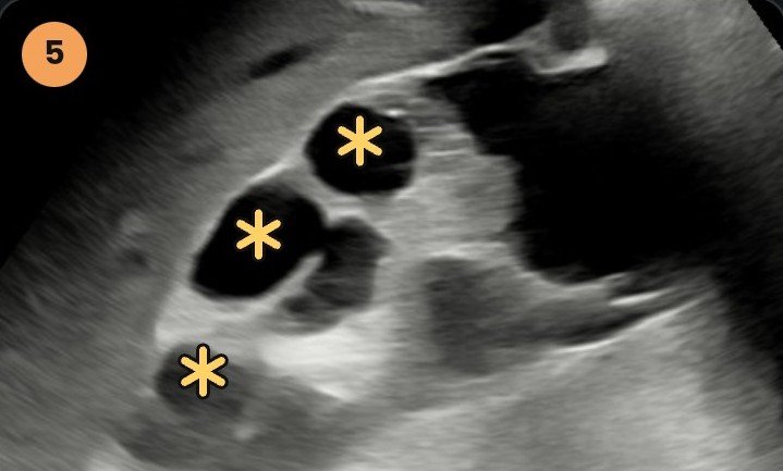

Acute SBO — dilated stomach, duodenum and jejunum with multiple jejunal diverticula; abrupt mid-jejunal transition with an anticlockwise mesenteric twist: volvulus. Normal SMA–SMV relation (no malrotation), no mass. Mild ascites; bilateral polycystic kidneys.
Segmental jejunal volvulus with multiple jejunal diverticula as the lead point
Management — resection of the diverticula-bearing segment with primary anastomosis; uneventful recovery.
Twist flagged on Day-0 Doppler; confirmed on CECT with cause and confirmed at surgery.
How the twist happens
The pouches
Mucosa bulges out where vessels pierce the mesenteric border.
The lead point
The pouch-laden loop becomes a lead point and rotates.
The whirl
Vessels spiral round the root; the bowel above it obstructs.
Kidney findings
USG: both kidneys normal in size, with multiple simple cortical cysts up to 30 mm, more on the right; no calculus or hydronephrosis. CECT: more than five cysts in each kidney, the largest 58 × 42 mm at the right lower pole.
Polycystic kidneys & diverticula
Colon — in kidney failure, diverticulitis struck 20% of ADPKD patients vs 3% of others (Lederman 2000); a US nationwide inpatient study found diverticulosis about twice as often (Duarte-Chavez 2021).
Small bowel — jejunal diverticula are reported in ADPKD, rarely, and can perforate (Morelle 2010; Naito 2024).
Why? — polycystin-1 and -2 are expressed in intestinal smooth muscle; a causal link is proposed, not yet proven (Morelle 2010).
Polycystic kidneys? Check the bowel for diverticula.