Baroda Road, Halol Mon–Sat 9 AM–7 PM · MRI, CT, X-ray, OPG and lab 24×7
Waveform Wonders · Interesting case · Head & Neck

Thyroglossal Fistula: Tracing the Tract on CT Sinogram and MRI

Midneck sinus → CT sinogram + MRI → the whole tract mapped

A neck sinus that keeps discharging? Trace the whole tract.

CTMRI Dr Abhishek Agarwal · 01 Nov 2026
Thyroglossal Fistula: Tracing the Tract on CT Sinogram and MRI
PatientPatient with a long-standing midneck sinus and occasional pus discharge
TopicHead & Neck
ImagingCT, MRI

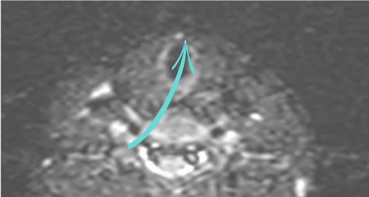
A long-standing sinus in the midline of the neck with occasional pus discharge. A CT sinogram traced the fistula up to the foramen caecum at the base of the tongue, and MRI showed the tract also reaching the thyroid isthmus, a part that contrast had not filled.

Case timeline

  1. Presentation

    Long-standing sinus in the midneck region with occasional pus discharge.

  2. CT sinogram

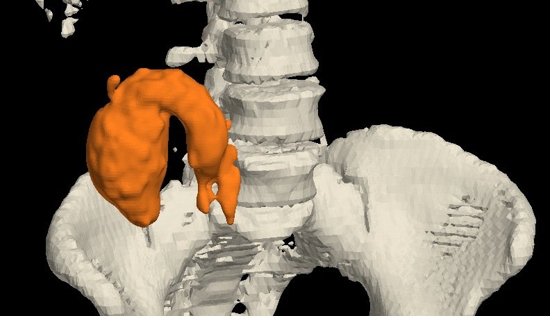
    Contrast injected through the track showed the fistula running up to the foramen caecum at the base of the tongue. 3D reconstruction showed the track clearly.

  3. MRI neck

    The track also extended to the thyroid isthmus, a part not opacified on CT.

Imaging findings

Clinical

External opening
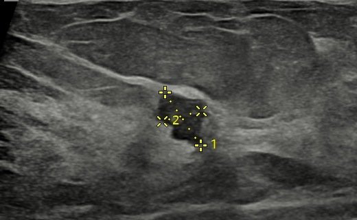
Midneck sinus: The external opening in the midline of the neck. 1
Midneck sinus The external opening in the midline of the neck.

CT sinogram

Contrast through the track · 3D and sagittal
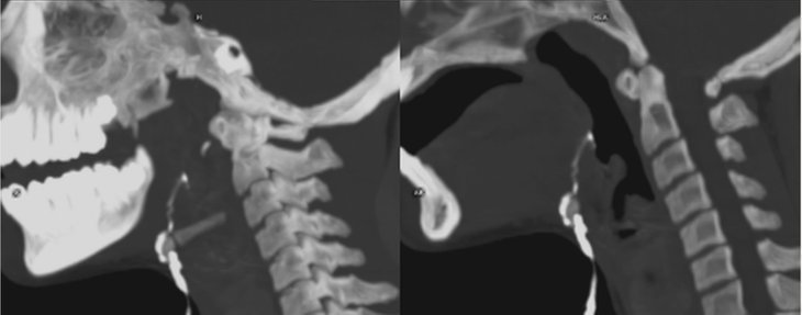
3D reconstruction: The contrast-filled track, shown clearly in 3D. 2
3D reconstruction The contrast-filled track, shown clearly in 3D.
Sagittal CT: Contrast in the track, running up towards the base of the tongue. 3
Sagittal CT Contrast in the track, running up towards the base of the tongue.

MRI neck

Track at the thyroid isthmus: MRI shows the track reaching the thyroid isthmus (arrow), a part not opacified on CT. 4
Track at the thyroid isthmus MRI shows the track reaching the thyroid isthmus (arrow), a part not opacified on CT.

Diagram

Course of the thyroglossal tract
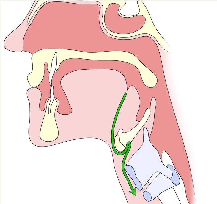
Course of a thyroglossal tract: From the foramen caecum of the tongue down the midline of the neck (arrow). 5
Course of a thyroglossal tract From the foramen caecum of the tongue down the midline of the neck (arrow).
CT + MRI

A fistulous track from the midneck opening up to the foramen caecum at the base of the tongue (CT sinogram), also extending to the thyroid isthmus (MRI): thyroglossal fistula.

Final diagnosis

Thyroglossal duct fistula

The CT sinogram mapped the track to the foramen caecum, and MRI added the part down to the thyroid isthmus that contrast had not filled.
Teaching points

What this case teaches

Take-home message

A neck sinus that keeps discharging? Trace it end to end: the CT sinogram shows the length, and MRI shows what contrast misses.

  1. Typical course — a thyroglossal duct fistula runs up to the foramen caecum of the tongue. It can be associated with a thyroglossal cyst.

  2. Discharging now? — MRI helps when there is active discharge from the external opening.

  3. Map the full length — a CT fistulogram (sinogram) defines the entire length of the tract.

  4. Use both — CT and MRI can complement each other: here, MRI showed the part that contrast did not fill.

Need a scan or an appointment?

We’re open Mon – Sat: 9:00 AM – 7:00 PM | Sun: By Appointment Only | 2D Echo: Thu 5–6 PM | MRI / CT / XRAY / OPG / Pathology: 24×7 available — walk in or call ahead.