Baroda Road, Halol Mon–Sat 9 AM–7 PM · MRI, CT, X-ray, OPG and lab 24×7
Waveform Wonders · Interesting case · Paediatrics

Hypertrophic Pyloric Stenosis in a 6-Week-Old

Thick pyloric muscle → gastric outlet obstruction → vomiting after feeds

Vomiting after every feed? Measure the pylorus.

UltrasoundX-ray Dr Abhishek Agarwal · 01 Nov 2026
Hypertrophic Pyloric Stenosis in a 6-Week-Old
PatientInfant, 1.5 months, persistent vomiting after each feed and poor weight gain
TopicPaediatrics
ImagingUltrasound, X-ray

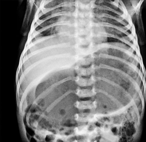
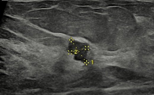
A 1.5-month-old baby had persistent vomiting after each feed and was not gaining weight. Ultrasound showed the typical pyloric wall thickening of hypertrophic pyloric stenosis, and an X-ray showed an over-distended stomach.

Case timeline

  1. Presentation

    Persistent vomiting after each feed and inability to gain weight.

  2. USG

    Typical pyloric wall thickening, suggesting hypertrophic pyloric stenosis.

  3. X-ray

    Over-distended stomach.

  4. Treatment

    Surgery: pyloromyotomy, in which the pyloric muscle is divided down to the submucosa.

Imaging findings

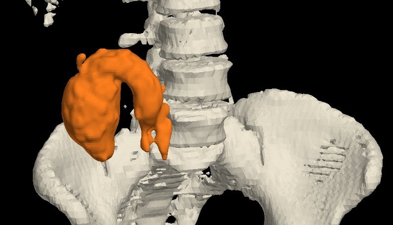
Thickened pylorus: Pyloric wall thickening with caliper readings (D1–D3); the inset outlines the hypertrophic pylorus. 1 Ultrasound
Thickened pylorus Pyloric wall thickening with caliper readings (D1–D3); the inset outlines the hypertrophic pylorus.
Over-distended stomach 2 X-ray
Over-distended stomach
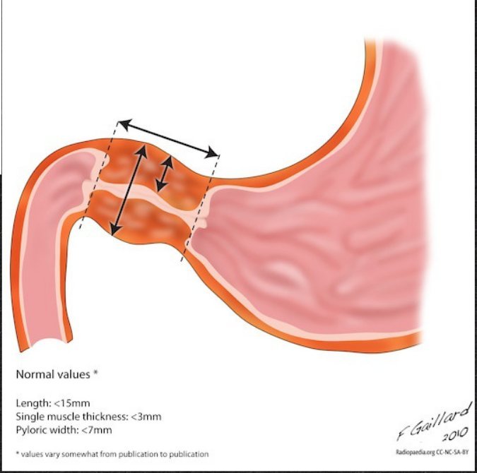
Pyloric stenosis: The thickened pyloric canal, with normal values. Illustration: F. Gaillard, Radiopaedia.org (CC BY-NC-SA). 3 Illustration
Pyloric stenosis The thickened pyloric canal, with normal values. Illustration: F. Gaillard, Radiopaedia.org (CC BY-NC-SA).
USG impression

Typical pyloric wall thickening: hypertrophic pyloric stenosis.

Final diagnosis

Hypertrophic pyloric stenosis

Treatment — pyloromyotomy, dividing the pyloric muscle down to the submucosa.
Teaching points

What this case teaches

Take-home message

Vomiting after every feed in a young infant? Measure the pylorus: a single muscle wall over 3 mm is the key number.

Ultrasound cut-offs

lengths in mm
>3

muscle thickness: a single wall on a transverse image (most accurate)

>15

pyloric length, measured longitudinally (>15–17 mm)

>1.5

pyloric volume, in cm³

>13

transverse diameter of the pylorus

  1. What it is — idiopathic thickening of the pyloric muscle, causing progressive gastric outlet obstruction.

  2. When — symptoms can start as early as 3 weeks, but it typically shows between 6 and 12 weeks of age.

  3. Classic picture — non-bilious projectile vomiting; the pylorus may be felt as an olive-sized mass in the right upper quadrant.

  4. Measure the muscle — a single muscle wall thicker than 3 mm on a transverse image is the most accurate sign.

Need a scan or an appointment?

We’re open Mon – Sat: 9:00 AM – 7:00 PM | Sun: By Appointment Only | 2D Echo: Thu 5–6 PM | MRI / CT / XRAY / OPG / Pathology: 24×7 available — walk in or call ahead.