A 30-year-old woman had knee pain and difficulty walking for a week after she started jogging. The X-ray raised the question of an osteoid osteoma in the medial tibia, but MRI showed a trabecular stress fracture of the medial tibial condyle, and the dense spots were incidental bone islands.
Case timeline
- Presentation
Knee pain with walking difficulty for a week after starting jogging.
- X-ray
Radiodense cortical lesion on the medial side of the tibia, suspicious for osteoid osteoma. MRI advised.
- MRI
Trabecular stress fracture of the medial tibial condyle: bone marrow oedema, and a thin hypointense line in the meta-diaphyseal region of the medial tibial plateau (yellow arrow).
- CT
A subtle dense line along the fracture line seen on MRI. The dense spots (*) were incidental bone islands.
- Treatment
Conservative: analgesia, ice, reduced weight-bearing and activity modification until the pain resolves.
Imaging findings
X-ray
 1
MRI
Marrow oedema · fracture line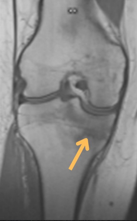
CT
Dense line · bone island

Reference chart
Stress fractures by site 6
Trabecular stress fracture of the medial tibial condyle: bone marrow oedema with a thin hypointense line. The X-ray “lesion” was an incidental bone island, not an osteoid osteoma.
Trabecular stress fracture of the medial tibial condyle
Treatment — conservative: analgesia, ice, reduced weight-bearing and activity modification until the pain resolves.
Address diet, vitamin D and calcium to prevent recurrence.