An adult patient came to the clinic with a broken edge on an upper front tooth, which left a notch in the smile. The tooth was rebuilt with a direct bonded composite restoration: tooth-coloured resin, shade-matched and bonded to the tooth, then shaped to its natural outline, with minimal removal of healthy tooth.
Case timeline
- Presentation
An adult patient with a broken edge on an upper front tooth, leaving a visible notch in the smile.
- Examination
Clinical photographs before treatment (A, C): the corner and part of the biting edge are missing.
- Treatment
Direct bonded composite restoration: tooth-coloured resin bonded to the tooth and built up to its natural outline.
- Result
The edge and outline are restored, and the repaired tooth blends with its neighbours (B, D).
Clinical photographs
Before and after
Cheek retractors in place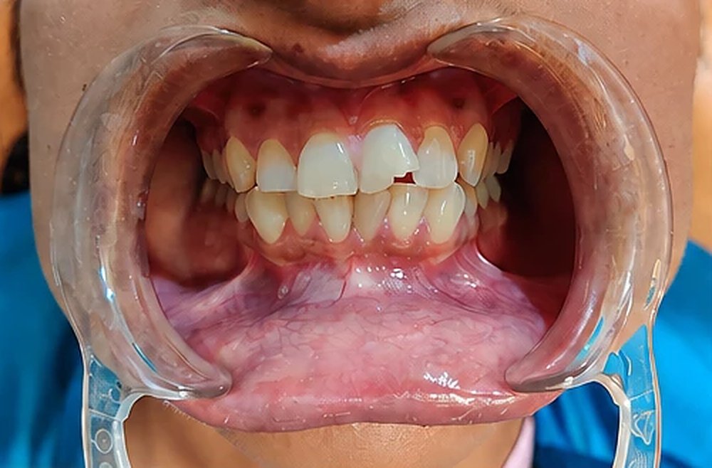
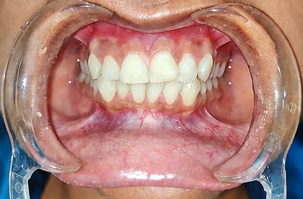
Look closer
The upper front teeth, enlarged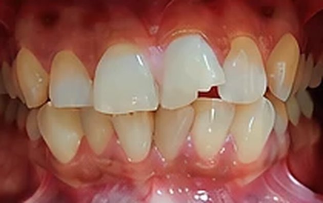 C
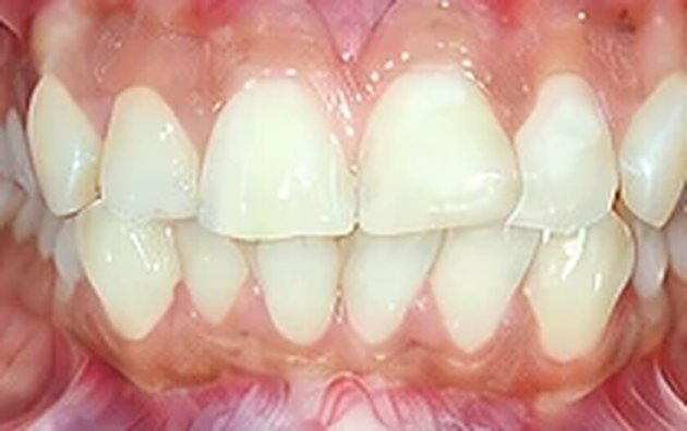 D
Composite bonding adds material to the tooth instead of cutting it down for a crown, so the healthy tooth underneath is kept.
Chipped (fractured) upper front tooth
Treated with a direct bonded composite restoration (tooth bonding).
How composite bonding is done
Shade and plan
The tooth is cleaned and checked, and a composite shade is matched to the neighbouring teeth.
Bond
The enamel is conditioned (etched) and a bonding agent applied, so the composite holds to the tooth with little or no drilling.
Build, shape, polish
Composite is added in thin layers, each hardened with a curing light, then shaped and polished to match the shine of natural enamel.
Why composite for a chipped tooth
Tooth-coloured: shade-matched, so the repair blends in.
Conservative: material is added to the tooth, so little or no healthy tooth is removed.
Repairable: a small chip in the composite later can usually be added to or polished, without starting again.
At the chair: usually done without waiting for a laboratory.
When a crown or veneer may suit better
A large break, a tooth that is already heavily filled or root-canal treated, or heavy grinding may need a stronger, laboratory-made crown or veneer.
If the broken tooth hurts or is sensitive, an X-ray (IOPA) comes first, because the nerve (pulp) may be involved.
Looking after a bonded tooth
Avoid biting nails, pens or ice with the repaired tooth, and wear a mouthguard for contact sports.
Tea, coffee and tobacco can stain composite over time; it can be polished again at routine check-ups.